
Medical anatomy education is changing with the development of digital technology and medical imaging. For many years, students mainly learned anatomy through textbooks, diagrams, and physical specimens. These methods remain valuable, but modern education requires more interactive ways to understand complex human structures.
The combination of medical images, three-dimensional reconstruction, and digital models creates new opportunities for anatomy teaching. The virtual human project CT scan approach represents this development by connecting clinical imaging data with educational applications, allowing students to explore anatomical structures in a more visual and interactive way.
The Connection Between CT Imaging and Digital Anatomy Learning
CT scanning provides detailed cross-sectional images of the human body. These images show internal structures layer by layer and are widely used in medical diagnosis. In anatomy education, CT data can help students understand how organs, tissues, and anatomical systems are arranged in real human bodies.
Traditional anatomy learning often requires students to imagine three-dimensional relationships from two-dimensional materials. Digital reconstruction changes this learning process by allowing structures to be viewed from different directions. Students can observe the same anatomical area through multiple perspectives and develop a clearer understanding of spatial relationships.
The development of the virtual human project CT scan concept allows medical educators to connect imaging information with three-dimensional learning environments. Instead of studying isolated images, students can explore reconstructed models that show the relationship between different structures.
This approach is especially useful for complicated regions such as the head, chest, and abdominal areas. These parts contain many connected structures that are difficult to understand through simple illustrations. Digital visualization provides additional support for students during anatomy courses.
Why Digital Human Models Are Important for Modern Education
Digital human models are not designed to replace traditional anatomy education. Instead, they provide an additional learning method that combines technology with established teaching practices. Teachers can use digital models to explain structures before students move to other practical learning activities.
Interactive systems allow users to adjust views, focus on specific structures, and compare different anatomical layers. This flexibility gives teachers more options when designing lessons. Students can also review important information repeatedly, which supports independent learning.
At DIGIHUMAN, we focus on developing digital anatomy solutions that connect medical data with teaching needs. Our work is based on the idea that accurate visualization can help educational institutions create more effective learning environments.
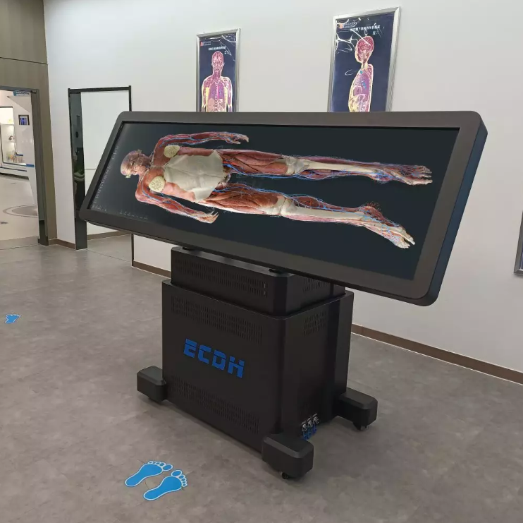
The HD Digihuman Virtual Anatomage Table applies real human body data reconstruction technology. The system combines traditional anatomy teaching with digital interaction. It includes CT and MRI image resources, allowing users to browse sequence images and review related case information, including imaging findings and imaging diagnosis.
Interactive Functions That Support Anatomy Teaching
One important advantage of digital anatomy platforms is the ability to interact with anatomical structures. In a traditional classroom, teachers usually explain structures through fixed images or physical models. Digital systems allow structures to be enlarged, reduced, rotated, and explored from different angles.
The HD Digihuman Virtual Anatomage Table allows users to manage anatomical information through various functions. Structures can be hidden, made transparent, displayed separately, marked, or collected for teaching purposes. These features help educators organize lessons based on different learning goals.
The system also supports multiple user accounts and customized presets. This allows different teaching groups to prepare suitable learning materials. For medical schools and training institutions, flexible content management can improve classroom organization.
The hardware design supports different teaching environments. The table uses an 88-inch display with 3840×1080 resolution, infrared touch operation, and an adjustable screen structure. The display can be lifted vertically and reversed 90 degrees according to teaching requirements. The product has CE and FCC certifications.
The Future Development of Anatomy Education
The future of anatomy education will continue to involve cooperation between medical knowledge and digital technology. As imaging technology develops, more educational resources can be created from real medical data. These resources can support different teaching methods and provide students with more opportunities to understand human structures.
The virtual human project CT scan approach shows how medical imaging can become part of a wider educational system. By combining CT images, MRI images, and three-dimensional reconstruction, digital anatomy platforms can create a learning process that connects theory, visualization, and practical understanding.
Digital tools are also becoming valuable for institutions that need scalable teaching resources. Universities, medical training centers, and healthcare education organizations can explore these solutions based on their own curriculum requirements.
Building New Possibilities for Anatomy Learning
Modern anatomy education is moving toward more interactive and data-based learning methods. Digital human models provide a bridge between medical imaging and classroom teaching, helping learners understand structures in a more visual way.
Through continuous development in digital anatomy technology, DIGIHUMAN aims to support professional education environments with practical solutions. The combination of accurate medical data and interactive learning tools creates new possibilities for anatomy teaching.
As education continues to evolve, the role of digital models will become increasingly connected with how students study human structures. The application of the virtual human project CT scan concept provides a valuable direction for future medical learning, while digital anatomy systems continue to support educators in building more flexible and informative teaching experiences.

MEDICA 2026 in Germany: What Medical Training Technology Looks Like This Year
Medical schools are being asked to train more people than they can teach. The World Health Organization's first annual health...

What Is Augmented Reality Medical Training
Medical schools have a quiet math problem. A class of thirty students often shares a single cadaver, and the numbers...

Hierarchical Structure of Anatomy: Understanding the Human Body From Systems to Detailed Structures
The human body is a complex organization built through multiple levels of structure. From large body systems to tiny anatomical...