
Medical schools have a quiet math problem. A class of thirty students often shares a single cadaver, and the numbers get worse outside major cities. That gap shapes everything about how anatomy is taught, from how long a lab session runs to how much a student actually gets to touch. It is also the reason augmented reality (AR) has moved out of demonstration booths and into real classrooms.
Why One Cadaver Is Never Enough for a Medical Class
A body can be dissected once. Once a structure is cut, it stays cut, and the next group of students inherits a specimen that has lost detail. Anatomy departments track the ratio of students to cadavers for this exact reason, because the ratio decides how much hands-on time any single learner gets.
The gap is not small. A survey of 55 medical colleges in mainland China found ratios running from 8.4 students per cadaver at top-ranked schools to 24 in western provinces, against a teaching benchmark of 4 to 7 (Chen et al., Anatomical Sciences Education, 2018).
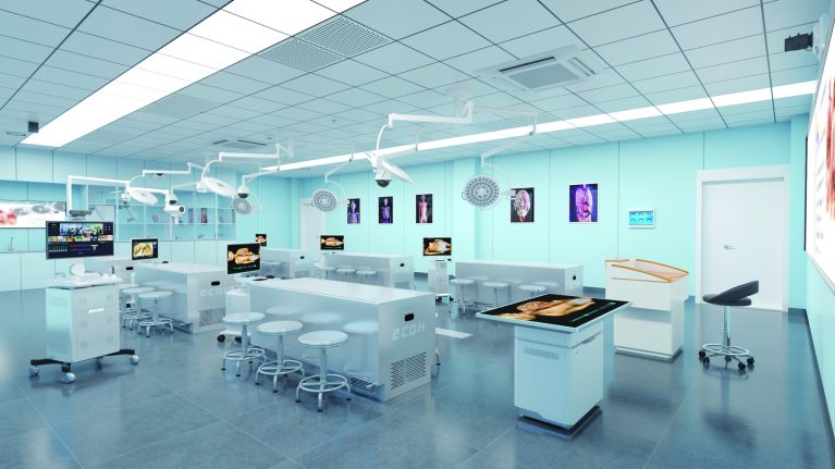
The shortage becomes visible the moment a class gathers. When thirty students crowd around one table, only a handful get to work with the material. The rest watch, take notes, and try to remember what they saw. Observation slowly replaces practice, and a student who never personally traces a nerve through surrounding tissue carries that uncertainty into clinical training.
A skilled teacher can point to a structure and explain its neighbors, and can slow a dissection down to walk the class through each step. What a teacher cannot do is multiply the specimen or hand every student their own.
Schools fill the gap with whatever tools they have. Charts, plastic models, and recorded lectures each cover part of the ground.
- Charts show structure without depth, so students struggle to judge how far one layer sits behind another.
- Plastic models hold a fixed shape but cannot be peeled apart, so what lies underneath stays hidden.
- Recorded dissections demonstrate a sequence but cannot be paused, rotated, or replayed from another angle.
Each tool answers a different question, and none of them lets a student repeat an action until it becomes automatic.
How Does Augmented Reality Help with Medical Purposes?
AR keeps the room you are standing in and adds anatomical structures on top of it. A student wears 3D tracking glasses with head tracking, holds an interactive pen, and works with a digital body that appears to occupy the same space as the physical table.
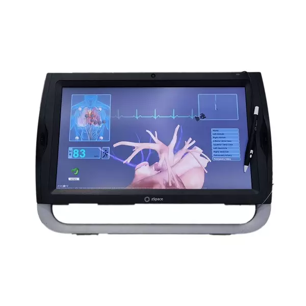
This is the practical difference between AR and virtual reality. VR replaces the room and asks the student to step into a headset. AR keeps the real environment visible and layers information over it, which matters when the skill being taught involves real instruments and real spatial judgment.
| Feature | Traditional Tools | VR | AR |
| Real classroom stays visible | Full | Blocked by headset | Full |
| Digitally reconstructed anatomy | Not available | Full 3D model | Layered on the room |
| Each student can repeat the task | One run only | Repeatable | Repeatable |
| Multiple viewers on one screen | Shared viewing | Limited to headset | Shared on screen |
Research backs the approach. A meta-analysis of 13 studies covering 654 participants found that AR training significantly improved performance time, confidence, and satisfaction compared with conventional teaching methods. Anyone asking how does augmented reality help with medical purposes will find the answer sitting in repetition.
How can augmented reality be incorporated in medical care? The classroom is the entry point, not the ceiling. The same setup supports clinical skills rehearsal, where trainees walk through a procedure from start to finish before they ever touch a patient. It supports group teaching, where one operator drives the session and an entire class follows on the screen.
It also supports regional anatomy work, where students compare a digital layer against a physical specimen on the same table. Schools that ask this question generally land on two tracks: self-directed study, where a student works through structures at their own pace, and instructor-led demonstration, where the teacher controls the sequence.
In practice, a typical session looks less dramatic than the marketing suggests. The sequence is short and repeatable:
- Put on the tracking glasses and pull up a region of the body.
- Turn the head to shift the viewing angle, the way you would lean around a real specimen.
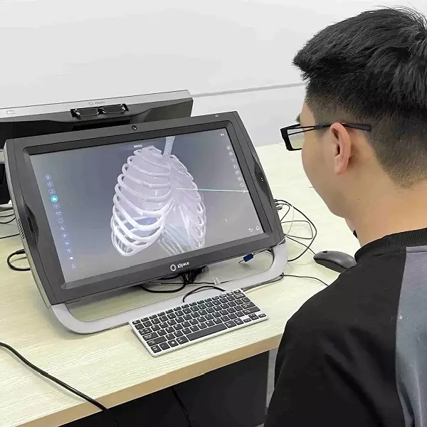
- Select a structure with the pen and isolate it from surrounding tissue.
- Remove a layer, see what sits beneath it, and put it back.
Steps three and four are the ones that matter. They repeat until the sequence is familiar rather than something the student has to think through.
Building an AR Anatomy Classroom: What It Takes
The technology only helps when the anatomy data underneath it is accurate. A digital body is useful only if every structure sits where it actually belongs, at a level of detail that survives close inspection. That constraint is what we work against at DIGIHUMAN, and why we have stayed in anatomy teaching since 2002. The system itself is built around a specific set of figures.
- 6,000+ anatomical structures rebuilt from continuous cross-sectional data taken from bodies free of organic disease and defects
- 5,000 digital models, covering both undergraduate and postgraduate teaching stages
- 1mm to 1mm sectional precision, with original tomographic images carrying up to 1.2 billion pixels
- 30 seconds or less to load the full system through our own Tai engine
- Every section sourced and vetted by the Committee of Experts of the Chinese Society for Anatomical Sciences
Customers in more than 40 countries use our systems in virtual anatomy laboratories, digital anatomy laboratories, and regional anatomy training centers. The same equipment serves medical schools, doctor training programs, and research institutions.
Conclusion: What It Means When Your School Is Considering AR
Cadavers will not become more plentiful, and the pressure to train more health workers keeps climbing. Every year, more students enter medical programs than the supply of donated bodies can support, and the gap lands on the anatomy lab.AR does not replace the specimen table, and it is not meant to. What it changes is how far a single specimen can reach. A structure that a student once watched someone else dissect becomes something they can open, examine, and close on their own.
That shift matters most for the students who currently get the least hands-on time, the ones at larger schools and in regions where specimens are scarcest. The technology keeps improving, and the underlying anatomy data keeps getting more detailed. What has not changed is the reason schools look at how can augmented reality be incorporated in medical care in the first place: they need a way to teach more students without waiting for a resource that will not arrive fast enough.

MEDICA 2026 in Germany: What Medical Training Technology Looks Like This Year
Medical schools are being asked to train more people than they can teach. The World Health Organization's first annual health...

Hierarchical Structure of Anatomy: Understanding the Human Body From Systems to Detailed Structures
The human body is a complex organization built through multiple levels of structure. From large body systems to tiny anatomical...

Virtual Human Project CT Scan: How Digital Human Models Support Modern Anatomy Education
Medical anatomy education is changing with the development of digital technology and medical imaging. For many years, students mainly learned...