
Understanding the relationship between airway structure and airflow is an important topic in medical research and education. The human lung contains a complex network of branching airways, where changes in shape and diameter can influence how air moves through different regions. With the development of medical imaging and computational methods, researchers can study these structures in more detail.
Modeling the bifurcating flow in a CT-scanned human lung airway has become a useful approach for connecting anatomical information with airflow analysis. This method allows researchers to examine how air travels through different airway branches and provides insights for respiratory studies, medical device development, and related applications.
Understanding Airway Bifurcation Through Medical Imaging Data
The respiratory system is formed by a series of branching pathways that begin with the trachea and continue into smaller bronchi and bronchioles. Each bifurcation creates a new flow environment, where airflow direction, velocity, and distribution may change according to airway geometry. Studying these structures helps researchers better understand the relationship between anatomy and respiratory function.
CT scanning provides detailed cross-sectional images of the human body, which can be reconstructed into three-dimensional anatomical models. These digital models allow researchers to observe airway shapes that are difficult to understand through traditional two-dimensional images. The reconstructed structures can show differences in airway length, diameter, and branching patterns.
When medical imaging data is combined with computational methods, researchers can create simulations based on realistic anatomical conditions. This approach provides a way to examine airflow behavior inside complex airway structures. Through modeling the bifurcating flow in a CT-scanned human lung airway, researchers can analyze how air moves through different branches and how anatomical features influence flow patterns.
How Computational Analysis Explains Airflow Changes Inside Airways
Computational analysis, including computational fluid dynamics (CFD), is commonly used to study fluid movement in complex biological structures. In airway research, CFD models use reconstructed anatomical data to simulate airflow under different conditions. These simulations can display changes in airflow velocity, pressure distribution, and movement direction.
The bifurcation area is an important part of airflow analysis because air does not simply move in a straight path. When airflow reaches a branching point, the geometry of the airway affects how the flow splits between the daughter branches. Factors such as airway angle, surface shape, and internal space can influence the movement of air.
By analyzing simulation results, researchers can compare airflow characteristics in different airway models. This information can support studies related to respiratory anatomy, biomedical engineering, and medical education. Digital analysis also helps students understand that anatomical structures are not isolated shapes but functional systems connected with physical processes.
Connecting Digital Anatomy With Medical Education
Modern medical education is gradually combining anatomical knowledge with digital technologies. Traditional anatomy learning often depends on physical specimens, textbooks, and static images. Digital visualization provides another learning method by allowing students to explore anatomical structures from different perspectives.
At DIGIHUMAN, we focus on developing digital anatomy solutions that connect real human data with interactive learning experiences. DIGIHUMAN uses reconstructed human body data to support the visualization of anatomical structures and medical imaging information. These technologies help educators present complex body systems in a more interactive format.
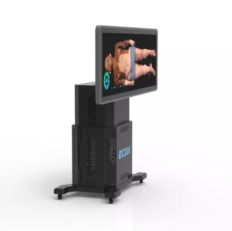
Our HD Digihuman Virtual Anatomage Table applies real human body data reconstruction and supports the display of CT and MRI sequence images. The system allows users to view three-dimensional structures, enlarge or reduce models, and rotate anatomical views from different angles. Different structures can also be marked, hidden, made transparent, separately displayed, or reviewed through selected functions.
For classroom and training environments, the table includes an display with a resolution of 3840×1080, infrared touch interaction, and adjustable screen positioning. The screen can be lifted, lowered, and adjusted according to different teaching requirements. With CE and FCC certifications, the system is designed for professional educational applications.
Supporting Future Learning With Integrated Anatomy Visualization
The combination of medical imaging and computational analysis creates new opportunities for understanding the human body. Instead of viewing anatomy only as fixed structures, students and researchers can explore how form and function influence each other. Airway studies are one example of how digital models can connect anatomical details with scientific analysis.
As medical education continues to develop, visualizing complex systems will become increasingly important. Interactive platforms can help learners examine anatomical relationships, review imaging information, and understand scientific concepts through direct observation. This approach creates a connection between clinical knowledge, research methods, and classroom learning.
Through modeling the bifurcating flow in a CT-scanned human lung airway, educators can introduce students to the relationship between airway anatomy and airflow simulation. We at DIGIHUMAN continue to explore digital solutions that support anatomy education by combining human data reconstruction, visualization technology, and interactive learning tools.
Bringing Anatomy and Analysis Together
The study of lung airflow demonstrates how anatomy and computational methods can work together. CT-based reconstruction provides the foundation for creating realistic models, while computational analysis helps explain how airflow interacts with these structures. This combination offers valuable references for medical research and teaching.
By integrating digital anatomy technologies into education, we aim to help institutions present complex anatomical information in a clearer way. Our solutions, including the HD Digihuman Virtual Anatomage Table, provide tools for exploring human structures and medical images. With this approach, modeling the bifurcating flow in a CT-scanned human lung airway becomes not only a research topic but also a learning opportunity that connects anatomy, technology, and medical knowledge. DIGIHUMAN supports this connection through digital visualization solutions designed for professional education environments.

MEDICA 2026 in Germany: What Medical Training Technology Looks Like This Year
Medical schools are being asked to train more people than they can teach. The World Health Organization's first annual health...

What Is Augmented Reality Medical Training
Medical schools have a quiet math problem. A class of thirty students often shares a single cadaver, and the numbers...

Hierarchical Structure of Anatomy: Understanding the Human Body From Systems to Detailed Structures
The human body is a complex organization built through multiple levels of structure. From large body systems to tiny anatomical...