
Medical simulation has become an essential part of healthcare education, allowing learners to develop practical skills before treating real patients. From traditional cadaver dissection to today’s virtual anatomy tables, simulation has evolved alongside advances in medical science and digital technology. So, why is medical simulation important? This article explores the answer and explains how digital anatomy platforms are reshaping anatomy learning.
The Growing Importance of Medical Simulation
Medical simulation is important because it creates realistic clinical experiences without exposing patients to unnecessary risk. It allows healthcare professionals to develop practical skills, refine decision-making, and prepare for challenging situations before applying those skills in real clinical settings.
Medical simulation has become increasingly important for several reasons. Modern healthcare involves more sophisticated technologies, complex procedures, and higher expectations for patient safety than ever before.
At the same time, healthcare professionals are expected to make accurate clinical decisions under pressure while working effectively within multidisciplinary teams. These challenges have made practical, experience-based training an essential part of professional development.
Medical simulation has continuously evolved alongside medical practice and digital technology. Early anatomy education relied primarily on cadaver dissection, giving learners direct experience with real human anatomy. Physical anatomical models later provided reusable teaching resources for classrooms, while mobile learning applications made anatomy more accessible beyond laboratories.
As computing power improved, virtual reality (VR) and augmented reality (AR) introduced immersive ways to explore anatomical structures and clinical scenarios. These innovations have expanded the possibilities of medical simulation, paving the way for more integrated digital anatomy platforms that combine visualization, interaction, and flexible teaching tools.
What Does Medical Simulation Do?
Medical simulation recreates realistic healthcare scenarios so learners can apply medical knowledge, practice clinical skills, and build confidence before treating real patients. Instead of simply teaching medical concepts, it supports simulation-based learning, allowing participants to learn by doing in a structured and supervised environment.
Medical simulation uses a variety of educational methods to reproduce clinical situations for teaching, training, and assessment. Depending on the learning objectives, it may involve standardized patients, task trainers, high-fidelity manikins, computer-based scenarios, or immersive digital technologies.
These tools encourage learners to assess patient conditions, make clinical decisions, perform interventions, and strengthen clinical reasoning—the ability to interpret information and choose appropriate actions in real clinical situations.
One of its most important roles is helping learners translate theory into practice. While textbooks and lectures provide the scientific foundation, simulation gives participants opportunities for deliberate practice, enabling them to repeat procedures, receive immediate feedback, and refine both technical and psychomotor skills before working with real patients.
Many simulation programs also include debriefing, a guided discussion after each scenario that helps learners reflect on their decisions, identify areas for improvement, and reinforce future performance.
Medical simulation also develops communication, teamwork, and clinical decision-making. Through interprofessional education (IPE), physicians, nurses, pharmacists, and other healthcare professionals can train together, improving role awareness and coordinated patient care in realistic scenarios.
Another major advantage is the ability to recreate low-frequency but high-risk clinical events, such as cardiac arrest, major trauma, medication errors, or rapidly deteriorating patient conditions. Repeated exposure to these scenarios helps healthcare teams respond more confidently and consistently when they occur in real practice.
Skills Developed Through Medical Simulation
- Transforming theoretical knowledge into practical clinical skills.
- Providing a safe environment for deliberate practice, feedback, and continuous improvement.
- Strengthening clinical reasoning, communication, and interprofessional collaboration.
- Preparing learners for routine care as well as rare, high-risk clinical events.
The Rise of Virtual Anatomy Tables in Modern Medical Education
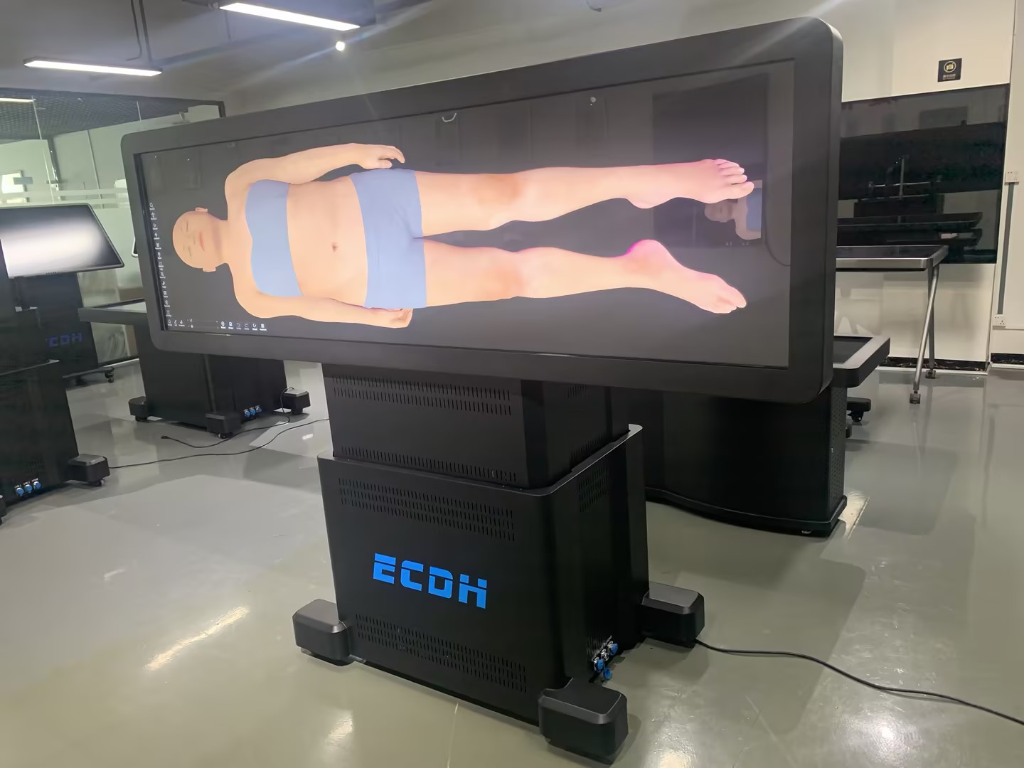
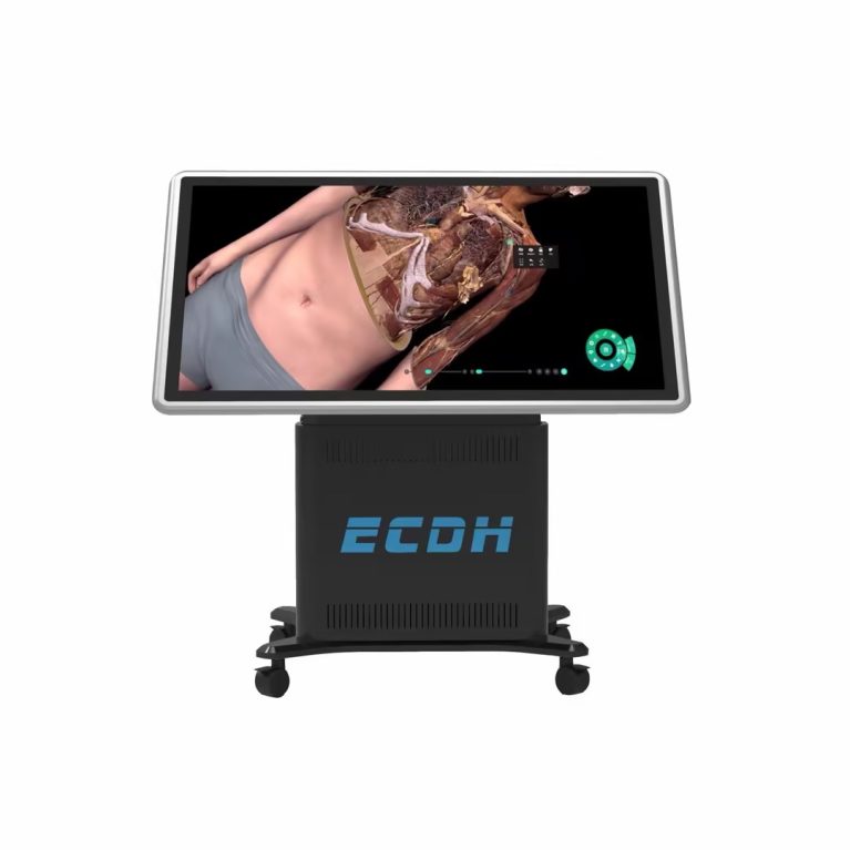
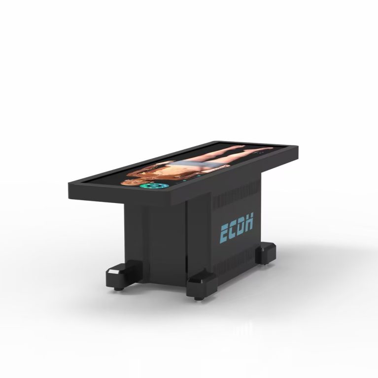
Virtual anatomy tables represent one of the latest developments in the evolution of anatomy education. As medical simulation has expanded beyond cadaver dissection, physical models, and standalone VR or AR applications, educators have increasingly adopted integrated digital anatomy platforms that combine visualization, interaction, and clinical learning in a single system.
How Does a Virtual Anatomy Table Work?
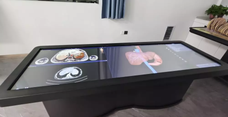
A Virtual Anatomy Table recreates the human body digitally by combining several core technologies. It is built on high-resolution digital human datasets derived from continuous cross-sectional images of real human bodies, which are reconstructed into accurate three-dimensional anatomical models. This allows users to explore anatomical structures from virtually any angle while preserving realistic spatial relationships.
Core Technology | How It Supports Learning |
Digital Human Data | Uses male and female digital human datasets, including 17,000+ male cross-sectional layers and 16,000+ female cross-sectional layers. |
3D Reconstruction | Reconstructs 3D anatomical structures while maintaining consistency with the original position and form. The system displays more than 6,000 anatomical structures across 9 body systems. |
Interactive Touch Interface | Supports touch operations such as rotating, enlarging, reducing, and moving anatomical structures. It also provides courseware, 100+ teaching videos, and over 1,800 exercises. |
CT & MRI Integration | Integrates clinical cases with CT/MRI images and reconstructed 3D structures. It includes more than 1,700 CT/MRI images for anatomy and clinical learning. |
How Are Virtual Anatomy Tables Changing Anatomy Education?
Virtual anatomy tables are changing anatomy education by making learning more interactive, repeatable, and collaborative. Instead of relying solely on scheduled laboratory sessions, instructors can demonstrate complex anatomical relationships dynamically, while students can manipulate structures themselves, review lessons repeatedly, and explore anatomy from multiple perspectives.
The differences between traditional anatomy teaching and virtual anatomy tables can be summarized as follows:
Traditional approach | Virtual Anatomy Tables |
Learning mainly occurs during scheduled laboratory sessions. | Anatomy can be explored repeatedly in classrooms or during self-study. |
Cadaver specimens are limited in availability and reuse. | Virtual dissections can be repeated without specimen limitations. |
Anatomy and medical imaging are often taught separately. | CT and MRI images can be viewed alongside anatomical models. |
Demonstrations are primarily instructor-led. | Students can actively manipulate and explore anatomical structures. |
How Virtual Anatomy Tables Are Being Used Around the World
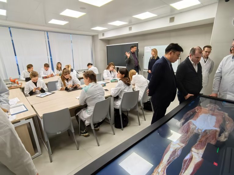
As digital anatomy becomes an increasingly important part of medical education, virtual anatomy tables are being adopted by medical schools, teaching hospitals, and healthcare training centers around the world. Today, the market includes solutions from Anatomage, BodyViz, Visible Body, and DIGIHUMAN, reflecting the growing demand for interactive anatomy teaching and digital learning resources.
Virtual anatomy tables are now used across a wide range of educational settings, including:
- Anatomy laboratories for undergraduate medical education
- Radiology and cross-sectional anatomy courses
- Surgical skills and procedure training
- Continuing medical education (CME) programs
- Clinical skills and interdisciplinary teaching sessions
Rather than replacing cadaver dissection, these systems complement traditional laboratory teaching by reinforcing anatomical concepts before and after hands-on dissection.
Among today’s digital anatomy solution providers, DIGIHUMAN develops digital anatomy platforms for medical schools, teaching hospitals, and healthcare training institutions. Our virtual anatomy table is built on real human digital datasets, featuring 17,000 layers of continuous anatomical sections, two high-definition male and female datasets, and 3D reconstruction with 0.1 mm precision.
These features allow users to explore anatomical structures with detailed anatomical visualization while supporting virtual dissection, DICOM-compatible CT and MRI image viewing, and interactive teaching activities.
Beyond technical capabilities, we have been adopted by numerous medical institutions across Mainland China and Hong Kong, including Peking University Health Science Center, Shandong University, and The University of Hong Kong. These examples reflect the growing role of virtual anatomy tables in modern medical education as institutions combine traditional anatomy teaching with digital learning resources to create more flexible and engaging educational experiences.
Conclusion
Medical simulation continues to reshape healthcare education by combining traditional teaching with digital technologies that make learning safer, more interactive, and easier to repeat. As virtual anatomy tables become a standard component of anatomy teaching, institutions are increasingly adopting digital platforms alongside cadaver-based instruction to prepare future healthcare professionals.
For institutions exploring digital anatomy solutions, DIGIHUMAN provides virtual anatomy tables designed to support anatomy teaching, medical imaging education, and collaborative learning in modern medical schools and teaching hospitals.

How Human Anatomy VR for Institutions Is Transforming the Modern VR Anatomy Teaching System
Human anatomy visualization has evolved from traditional methods to more digital and interactive approaches. Today, Human Anatomy VR for institutions provides...

3D Printed Anatomical Model Explained: Concept, Applications, and Advantages
Advances in 3D printing and digital modeling are reshaping many fields. One of the most visible changes can be seen...
Innovative Applications of Virtual Dissection Tables
The rise of the virtual dissection table has transformed how anatomy is taught in medical institutions. Traditional dissection has long...