
Human anatomy visualization has evolved from traditional methods to more digital and interactive approaches. Today, Human Anatomy VR for institutions provides organizations with new ways to explore anatomical structures through modern VR Anatomy Teaching Systems. By combining virtual environments with digital human technologies, these systems offer more flexible approaches for anatomy teaching, training, and research. This article examines the development of Anatomy Teaching Systems and how VR-based technologies are changing the way human structures are visualized and explored.
From Traditional Anatomy Learning to Digital Solutions: The Evolution of the Anatomy Teaching System
Anatomy education has gradually developed from physical observation to digital exploration, with technologies such as 3D modeling, virtual reality (VR), and augmented reality (AR) changing how institutions teach and learn human anatomy. These technologies are helping medical schools, healthcare organizations, and educational institutions create more flexible anatomy learning environments.
For many years, anatomy education depended mainly on cadaver dissection, physical models, and two-dimensional textbook illustrations. Cadaver-based learning remains an important part of medical training because it allows students to observe real human structures and understand natural variations. However, traditional approaches also involve practical challenges, including limited specimen availability, laboratory requirements, and the difficulty of providing repeated access for large groups of learners.
As medical education continues to expand, institutions are looking for additional tools that can support anatomy learning without being restricted by physical resources. Digital anatomy solutions have emerged as a way to provide repeatable and interactive learning experiences.
The development of anatomy teaching technology can be divided into several stages:
Development Stage | Learning Approach | Main Characteristics |
Traditional anatomy | Cadaver dissection | Direct observation of real human structures |
Visual learning | Textbooks and diagrams | Learning through 2D illustrations |
Digital anatomy | 3D models and software platforms | Interactive visualization of anatomical structures |
Immersive anatomy | VR Anatomy Teaching System | Exploration of anatomy through virtual environments |
These developments have created new possibilities for institutions that need flexible and repeatable anatomy learning resources. Instead of viewing a fixed image, learners can examine anatomical relationships from different angles and develop a clearer understanding of spatial connections.
Among these digital solutions, Human Anatomy VR for institutions represents a new direction in the development of modern anatomy teaching approaches.
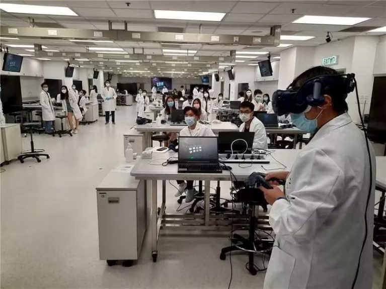
What Can Human Anatomy VR for Institutions Provide for Medical Training and Research?
Human Anatomy VR for institutions provides a digital environment where students and professionals can explore human body structures through detailed three-dimensional models. This approach helps learners intuitively grasp complex spatial relationships, depth, and structural proportions within the human body.
Centering on human biology, institutional VR systems provide comprehensive structural exploration across all anatomical levels, from whole-body systems to individual organs and tissues:
- Comprehensive anatomical visualization:Users can explore major body systems, such as the skeletal, muscular, nervous, and cardiovascular systems, while understanding how different structures are connected.
- 360-degree spatial observation:Three-dimensional models allow learners to view anatomical structures from different angles, isolate specific components, and better understand their positions and relationships within the body.
- Detailed structure examination:Digital anatomy platforms can support close examination of smaller anatomical components, helping learners study tissues and other detailed structures that are difficult to visualize through traditional materials.
- Medical imaging integration:Some VR anatomy solutions can incorporate imaging resources such as CT and MRI data to help learners connect anatomical models with clinical visualization methods.
Human Anatomy VR for institutions supports diverse application scenarios, including medical education for student structural learning, healthcare training for professional development, teaching demonstrations for dynamic instructional delivery, and scientific education for public knowledge dissemination.
These applications allow institutions to expand anatomy learning beyond traditional classroom settings and provide additional resources for different learning and research requirements across various educational environments.
These capabilities make VR anatomy systems suitable for institutional applications such as education, training, and research.
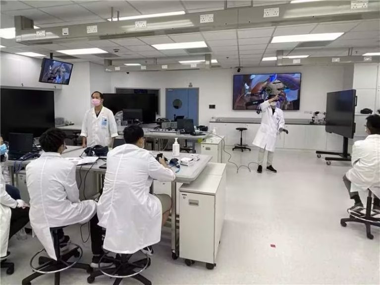
How Does a VR Anatomy Teaching System Create an Interactive Anatomy Experience?
Through VR headsets and hand controllers, users can interact with digital human models and adjust the virtual learning environment according to different anatomy topics. These operations allow learners to examine structures more flexibly and focus on specific areas during study sessions.
Unlike traditional anatomy materials that usually provide a fixed viewing angle, VR-based systems allow learners to actively control their observation process. For example, students can move around a virtual model, examine the relationship between surrounding structures, and switch between different anatomical views based on the learning objectives. This hands-on interaction helps transform anatomy learning from simple observation into a more active exploration process.
Common interactive functions in VR anatomy learning include:
- Rotation and movement:viewing anatomical structures from multiple directions to understand their spatial relationships.
- Zooming:examining smaller or complex structures that may be difficult to observe in traditional images.
- Showing and hiding structures:focusing on specific organs, systems, or tissues during learning.
- Separating anatomical components:studying the connections between different anatomical structures.
These interactive functions help learners build a clearer understanding of complex anatomical relationships. In addition, digital anatomy resources can be accessed repeatedly during different teaching sessions, providing institutions with a consistent learning environment.
Digital anatomy resources can also reduce dependence on physical specimens and provide a controlled learning environment for repeated practice.
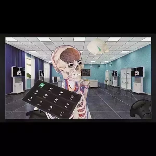
How DIGIHUMAN VR Anatomy Teaching System Supports Institutional Anatomy Applications
DIGIHUMAN VR Anatomy Teaching System is designed as a complete virtual anatomy teaching solution that combines 3D reconstruction, VR hardware, and multiple teaching modules to support anatomy education in institutions.
The system integrates multiple functional modules that work together within a single virtual anatomy environment. These include the wizard system, multimedia system, graphics and image system, voice system, puzzle system, practice system, UI system, and interactive system. Together, these modules support anatomical visualization, interactive exploration, learning activities, and classroom demonstrations.
The system provides several functions designed for classroom teaching and independent learning:
Product Feature | Function |
Wizard system | Guides first-time users through text and voice instructions |
Graphics and image system | Supports anatomical visualization through slice images, CT data, and other image resources |
Practice system | Provides exercises, including single-choice and multiple-choice questions, to review learning results |
Puzzle system | Allows users to assemble models in a 3D environment with different difficulty levels |
Multimedia system | Provides video-based teaching resources |
Interactive system | Supports access to different anatomical models |
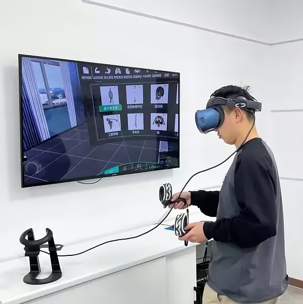
In addition to software functions, DIGIHUMAN VR Anatomy Teaching System uses VR components including VR headsets and handheld controllers. The equipment creates an immersive learning environment, while controllers allow users to operate virtual anatomy models during teaching activities.
The system also supports different presentation methods for institutional use. In addition to individual VR learning experiences, the virtual content can be displayed through connected screens, allowing teachers to share anatomy demonstrations with a larger group of learners.
Conclusion: Why Will Human Anatomy VR Continue to Matter in Modern Anatomy Teaching Systems?
Human Anatomy VR for institutions is becoming an important part of modern Anatomy Teaching Systems because it provides additional resources for anatomy learning, training, and research. By supporting digital visualization and repeatable learning, VR technology helps institutions create more flexible approaches to anatomy education.
Our DIGIHUMAN VR Anatomy Teaching System combines 3D reconstruction with dedicated educational software to support immersive learning. For institutions exploring modern digital anatomy options, contact our team to see how we can support your curriculum.

Why Is Medical Simulation Important? From Cadaver Dissection to Virtual Anatomy Tables
Medical simulation has become an essential part of healthcare education, allowing learners to develop practical skills before treating real patients....

3D Printed Anatomical Model Explained: Concept, Applications, and Advantages
Advances in 3D printing and digital modeling are reshaping many fields. One of the most visible changes can be seen...
Innovative Applications of Virtual Dissection Tables
The rise of the virtual dissection table has transformed how anatomy is taught in medical institutions. Traditional dissection has long...