
When pet owners hear that a dog may need an MRI, one of the first questions they often ask is: what does an MRI scan show on a dog? Magnetic resonance imaging can provide highly detailed views of internal structures, especially soft tissues that may be difficult to evaluate with conventional X-rays.
At DIGIHUMAN, we are interested in medical imaging and three-dimensional visualization because these technologies can make complicated anatomical relationships easier to understand. Although MRI is widely used in veterinary diagnosis, understanding what the images actually represent can help us appreciate how modern imaging supports the study of anatomy.
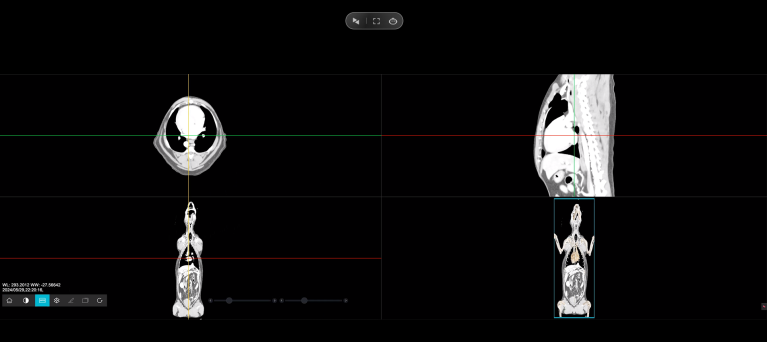
How Does MRI Work in Dogs?
MRI uses a strong magnetic field, radiofrequency signals, and computer processing to create detailed images of structures inside the body. Unlike X-ray imaging, MRI does not use ionizing radiation. The technique is particularly sensitive to differences in soft tissues and their water content.
During an examination, the dog is positioned inside the MRI scanner. Because movement can seriously affect image quality, veterinary patients generally need anesthesia or carefully managed sedation so that they remain still during the examination.
The resulting images can be viewed in different planes, allowing veterinary specialists to examine internal anatomy from multiple directions. This is especially valuable when structures overlap on a conventional two-dimensional image.
What Can an MRI Show in a Dog’s Brain?
The brain is one of the areas where MRI is particularly useful. Veterinary MRI can provide detailed images of the brain and help specialists investigate conditions involving the brain and surrounding structures.
For example, MRI may help evaluate suspected brain tumors, inflammatory conditions, structural abnormalities, and other neurological problems. It can also provide information about the tissues surrounding the brain that may not be sufficiently characterized by other imaging techniques.
This ability to visualize soft tissue in detail is one of the major reasons MRI has become an important diagnostic tool in veterinary medicine.
What Does MRI Show in a Dog’s Spine?
The spinal cord and spine are another major application of MRI. When a dog develops neurological symptoms such as weakness, difficulty walking, or other signs associated with the nervous system, a veterinarian may consider advanced imaging to investigate the underlying cause.
MRI can show the spinal cord, spinal canal, intervertebral discs, surrounding soft tissues, and related anatomical structures. It can therefore help identify problems such as disc herniation, spinal cord disease, inflammation, tumors, or narrowing of the spinal canal.
Rather than looking at one isolated structure, specialists can examine how different tissues relate to each other. This spatial information can be important when determining where an abnormality is located.
Can MRI Show a Dog’s Muscles and Joints?
Yes. MRI is particularly useful for evaluating many soft tissues, including muscles, ligaments, tendons, and structures associated with joints.
This can be valuable when a dog has persistent lameness, suspected soft-tissue injury, or a condition that is not adequately explained by initial examinations. MRI can provide detailed information about tissue structure and help veterinarians distinguish between different possible causes.
MRI can also provide information about the internal structure of bones, although other imaging methods may be more appropriate for certain types of bone abnormalities. The choice of imaging technique depends on the clinical question and the part of the body being examined.
What Can MRI Reveal About a Dog’s Abdomen?
MRI can also produce images of organs and soft tissues in other parts of the body. Veterinary sources note that MRI may be used to examine abdominal soft tissues as well as the brain, spinal cord, and other structures.
However, MRI is not automatically the best imaging method for every condition. X-rays, ultrasound, CT, and other diagnostic techniques each have different strengths. Veterinary professionals select an imaging method according to the suspected disease, anatomical region, clinical signs, and information required.
Therefore, asking what does an MRI scan show on a dog is really a question about both anatomy and imaging technology. An MRI does not simply create a photograph of a dog. Instead, it generates detailed images that allow specialists to investigate particular tissues and anatomical relationships.
Why Is Three-Dimensional Visualization Important?
Anatomy is inherently three-dimensional, and understanding how structures relate to one another is essential in medical education. DIGIHUMAN‘s Virtual Anatomy Table supports this learning process through systematic, regional, and sectional views of more than 6,000 three-dimensional anatomical structures.
The system also includes dissection videos, anatomy animations, and assessment resources. Although designed primarily for human anatomy education, these tools demonstrate how interactive 3D visualization can help learners explore complex anatomical structures and understand their spatial relationships.
Conclusion: Understanding What an MRI Shows in Dogs
An MRI scan can reveal detailed images of a dog’s brain, spinal cord, muscles, joints, and other soft tissues. This information helps veterinarians investigate conditions that may be difficult to assess using conventional imaging, although the most appropriate imaging method depends on the dog’s symptoms and suspected condition.
At DIGIHUMAN, we use digital anatomy visualization to make complex anatomical structures easier to explore and understand. While our Virtual Anatomy Table is designed primarily for human anatomy education rather than veterinary diagnosis, it reflects our commitment to making anatomy learning more visual, interactive, and accessible.

Cadaver vs. Virtual Anatomy: How PA Students Learn the Human Body
Two routes now run through every physician assistant anatomy course. One starts with a donor on a steel table, the...

How Are Human and Rat Anatomy Similar?
When we study anatomy, one of the most interesting questions we encounter is how are human and rat anatomy similar?...

What Is the Largest Organ in the Rat’s Anatomy?
When we study animal anatomy, one of the simplest questions can lead to a surprisingly interesting discussion: what is the...