
Two routes now run through every physician assistant anatomy course. One starts with a donor on a steel table, the other with a touchscreen and a dataset built from real human sections. The disagreement over cadaver vs virtual anatomy teaching methods for physician assistant students is really about what each route teaches and what a program can sustain. The comparison matters most where it is least visible, in what a graduate can do on the first day of clinicals.
The Clinical Essentiality and Evolving Challenges of Cadaver Dissection
Cadaver dissection has reshaped medical training, from its historical roots to modern practice, anchoring both clinical care and PA education. The roots run back to 1543, when Andreas Vesalius published De Humani Corporis Fabrica and replaced a millennium of textbook authority with what the dissection table showed. The method held as it worked, and generations of physicians have since learned the layout of the body, its variations and its three-dimensional relationships the same way, by taking it apart. For PA students, that first pass through a human body sets the reference every later clinical judgment is measured against.
Hands-on engagement with donors helps illuminate core anatomy and its clinical relevance. Tissue has temperature, resistance and variation, and a structure that looked straightforward in an atlas turns out to sit at an angle, to be absent, or to be doubled. It also builds essential, cross-disciplinary competencies required across modern healthcare settings: sustained concentration at a shared table, teamwork under time pressure, and the professional habit of treating a body as someone’s first patient rather than as material.
Widespread donor shortages, however, compounded by mounting facility costs and logistical burdens, are compelling medical programs to adopt alternative teaching methods alongside dissection, giving rise to more flexible, multimodal approaches to anatomy education. Donation is voluntary, regional and uneven, and a single donor supports a limited number of students before the preparation is spent. Around the specimen sit costs that never appear on a syllabus: procurement, transport, refrigeration, formaldehyde handling, ventilation and eventual disposal.
Virtual Anatomy: What a Dissection Table Adds
In response, medical education has resorted to interactive dissection tables and newer digital formats, aiming to convert anatomy into interactive datasets, by which trainees are allowed to isolate deep-seated structures beyond the capacity of a fixed donor. The data comes from real human cross-sectional images reconstructed in 3D, and a 3d virtual cadaver dissection table can open any plane of that reconstruction, in any order, as many times as a lesson requires. A structure buried behind bone stops being a problem of access and becomes a problem of sequence.
Such modalities have since emerged and transformed anatomical training, with unlimited repeat practice and clearer spatial orientation. A student who missed a relationship the first time can rebuild it in minutes instead of waiting for the next donor, and a structure that looked flat in an atlas can be rotated and re-sectioned from every angle. The table also removes the preservative: no formaldehyde exposure, no specimen decay, and no ethical weight attached to a mistake made during practice.
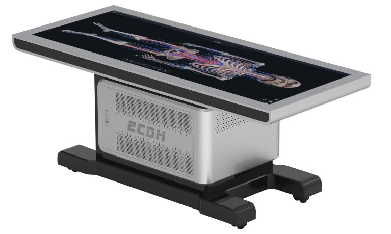
Still, no screen can replicate how human tissue feels in real conditions. A screen cannot tell a student that a structure is firmer than it looks, or that an artery twangs when stretched, and those are the cues a surgeon’s fingers depend on later. The honest reading is that the table reproduces the anatomy, not the encounter. A student who has already sectioned a region on screen arrives at the donor with a map, and the hour at the table goes into variation and texture instead of orientation.
Where the Two Diverge and Where They Meet
Cadaveric dissection demands extensive facility infrastructure, rigorous maintenance, and continuous donor supply, all of which affect how a curriculum is built and budgeted. The infrastructure is specific: a dedicated lab, ventilation built for preservatives, refrigeration for storage, and a donor program renewed every term. The comparison behind cadaver and virtual anatomy teaching methods for physician assistant students comes down to what a program can sustain.
Digital anatomy platforms, in comparison, prioritize scalable access, software versatility, and repeatable 3D spatial exploration, imposing minimal recurring costs. Access scales with a license rather than a donation, the software is updated rather than re-prepared, and the same model serves the next cohort unchanged. A 3D virtual cadaver dissection table can be sectioned by an entire class without being consumed. Set side by side, the two look like this.
| Dimensions | Cadaver dissection | Virtual anatomy table |
| Facility and supply | Dedicated lab, continuous donor program | Screen and software, no specimen required |
| Repeat practice | One donor, one preparation | Unlimited, any plane, any order |
| Recurring cost | Procurement, storage, disposal | Licenses and software updates |
| Tactile feedback | Real tissue under the fingers | Not reproduced on screen |
Despite the differences, however, both reinforce each other and boost student learning. Digital tools enable in-depth spatial comprehension, while hands-on dissection facilitates tactile and structural retention. Leveraging both optimizes pedagogical efficacy. The digital side builds the map: where a structure sits, what it crosses, and how it changes from section to section. The donor supplies the sense of resistance and variation that turns the map into something usable in a living patient. Programs that run both are not splitting the difference. They teach two kinds of knowledge, and each one makes the other hold.
Building an Integrated Anatomy Program
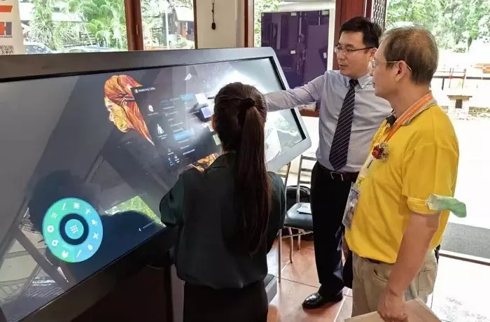
Virtual anatomy tables can be used as a valuable additive tool to traditional cadaveric dissection in modern anatomy education. Today, medical institutions worldwide are increasingly integrating digital platforms into traditional labs, in order to enhance spatial comprehension and curricular versatility.
DIGIHUMAN, a leading Chinese innovator in digital human research, equips anatomy departments across more than 40 countries, leveraging ultra-high-definition real human data—which has gained widespread academic recognition and global adoption—to power its flagship virtual anatomy tables.
- Data: continuous real specimen sections between 0.1 mm and 1 mm thick, with the original tomographic images reproduced at up to 1.2 billion pixels.
- Imaging: more than 1,700 corresponding CT and MRI images, so the gross specimen and the scan of the same region can be read against each other.
- Validation: every section vetted by the committee of experts of the Chinese Society for Anatomical Sciences, and the HD virtual dissection table system appraised by the Society in January 2021 at an international advanced level.
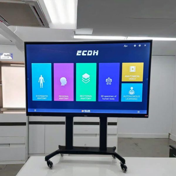
DIGIHUMAN’s range covers standard, HD, and mini tables plus 55, 86, and 98-inch teaching terminals, and the labs employing both modalities are already built that way. A specimen refrigeration dissection table handles the hands-on work, while a mini virtual table beside it runs the same region, so a class can move between the donor and the screen without changing rooms.
The Future of PA Anatomy Training
Cadaver and virtual anatomy vary in tactile experience, structural variability, and digital precision. Each has its own advantages and can be used in multiple scenarios. Integrating both points to a world where anatomy education delivers maximum diagnostic efficiency and well-rounded clinical competence.
For a PA student, the sequence is already visible: palpate the donor in the morning, section the same structure on screen in the afternoon, and carry both into the clinic.

How Are Human and Rat Anatomy Similar?
When we study anatomy, one of the most interesting questions we encounter is how are human and rat anatomy similar?...

What Is the Largest Organ in the Rat’s Anatomy?
When we study animal anatomy, one of the simplest questions can lead to a surprisingly interesting discussion: what is the...

What Is Fish Anatomy? A Simple Guide to How Fish Are Built and How They Survive
When we look at a fish swimming through water, its streamlined body may seem simple. However, beneath the scales and...