
Understanding the architecture of the human body requires a structured framework. Systemic anatomy serves as a core branch of anatomical science that organizes the body into distinct functional systems, allowing students and medical professionals to study how individual organs collaborate to sustain life.
The study of systemic anatomy has evolved over thousands of years. Early anatomical understandings date back to ancient Egypt and Greece, where pioneers like Galen began dissecting animals to deduce human organ functions. During the Renaissance, pioneers like Andreas Vesalius transformed the discipline through precise human dissection and detailed illustration, shifting medical education from philosophical speculation to direct observation. Over time, anatomists moved from examining isolated body parts to categorizing the body into organized, interconnected functional systems.
Below, we explore the core principles of systemic anatomy, the organ systems studied in medical curricula, and how technology is revolutionizing modern anatomical education.
What Is Systemic Anatomy?
Systemic anatomy is a specialized sub-field of gross anatomy that categorizes the human body based on specific physiological functions. Rather than focusing on a single region (such as the arm or thorax), systemic anatomy groups related organs and tissues into functional units—such as the digestive or nervous system—to study their structural organization and cooperative functions throughout the entire body.
On the one hand, in systemic anatomy, you will learn specialized tissues form organs, which serve as functional building blocks. On the other hand, systemic anatomy also integrate organs working toward a shared primary objective as a single system, and learn each the function of each system.

Core Systems in Medical Education and Research
In standard medical and biological education, systemic anatomy typically categorizes the human body into nine primary functional systems. Depending on the textbook or curriculum, some sub-disciplines isolate additional categories like the Skeletal System, Integumentary System (skin, hair, nails), or Cardiovascular System. The Primary Organ Systems include:
- Locomotor System (Musculoskeletal System): Combines the skeletal system (bones, joints, cartilage) and muscular system to provide structural support, protection for internal organs, and body movement.
- Digestive System: Comprises the oral cavity, esophagus, stomach, intestines, liver, and pancreas to process food, absorb nutrients, and eliminate solid waste.
- Respiratory System: Includes the nasal passages, larynx, trachea, bronchi, and lungs, facilitating gas exchange between the body and the external environment.
- Urinary System: Consists of the kidneys, ureters, bladder, and urethra, responsible for filtering blood, regulating fluid balance, and excreting liquid waste.
- Reproductive System: Encompasses male and female reproductive organs responsible for gamete production, hormone regulation, and continuation of the species.
- Endocrine System: Comprises hormone-secreting glands (e.g., pituitary, thyroid, adrenal) that regulate metabolism, growth, and cellular activity throughout the body.
- Circulatory System: Includes the cardiovascular system (heart, blood vessels, blood) and lymphatic system, delivering oxygen and nutrients while supporting immune defense.
- Sensory Organs: Specialized structures (eyes, ears, taste buds, olfactory receptors, skin sensors) that receive environmental stimuli and translate them into neural signals.
- Nervous System: Comprises the central nervous system (brain and spinal cord) and peripheral nervous system, serving as the master control unit for sensory processing, motor output, and internal coordination.
While each system performs a unique function, none operates in isolation. For example, the circulatory system transports hormones produced by the endocrine system, while the respiratory system relies on nervous system signaling to maintain breathing rates. Furthermore, principles of human systemic anatomy apply directly to comparative and veterinary anatomy. Because vertebrate species share homologous organ systems, learning human systemic anatomy provides a foundational understanding that extends across mammalian research and veterinary medicine.
How Medical Students Learn Systemic Anatomy
The methods used to teach systemic anatomy have undergone a dramatic transformation over recent decades.
Historically, traditional learning relied heavily on static 2D textbook diagrams, lectures, physical plastic models, and donor cadaver dissection. While cadaver dissection remains a valuable standard for spatial orientation, it presents challenges regarding facility costs, tissue preservation, and limited availability.
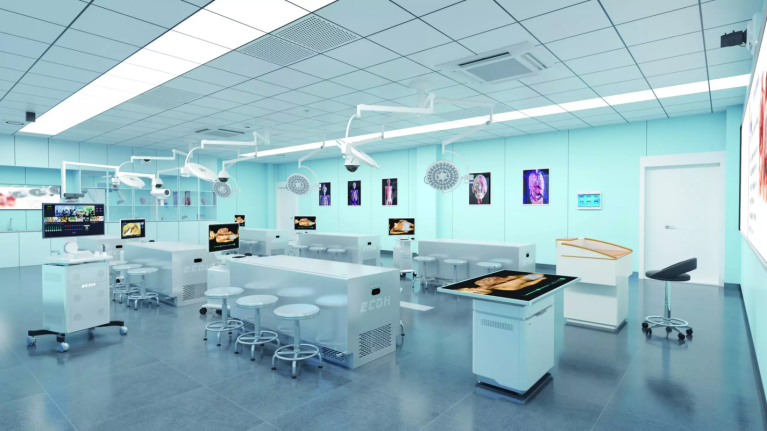
Today, digital technologies supplement traditional methods. Students utilize 3D interactive software, augmented reality (AR), virtual reality (VR), and full-scale Virtual Anatomy Tables (such as the Anatomage Table).
These digital 3D tools allow students to isolate individual organ systems, rotate models 360 degrees, and view cross-sections in real time. Virtual anatomy platforms enable endless digital dissections, allowing students to slice, undo, and re-examine organ structures layer by layer. Moreover, institutions can deliver standardized, highly accessible anatomy instruction without the continuous logistical demands of cadaver lab maintenance.
Conclusion
Systemic anatomy provides an organized, system-by-system approach to understanding human structure and function. By examining how individual organs align within broader physiological units, students build a foundational understanding necessary for clinical diagnostics, pathology, and surgery. Combined with modern 3D visualization technology, systemic anatomy education continues to become more intuitive, accessible, and impactful for future healthcare professionals.
At DIGIHUMANwe are proud to offer the Digihuman Virtual Anatomy Table as a transformative learning tool that enhances the educational experience for both instructors and students. Together, we can revolutionize anatomy education, making it more effective and engaging while preparing future healthcare professionals for success. Let’s redefine how systemic anatomy is taught, making mastery of this essential subject not only possible but also appealing.
FAQ
Q1: Comparison: Systemic Anatomy vs. Regional Anatomy vs. Physiology
A1: Here are the differences:
Feature | Systemic Anatomy | Regional Anatomy | Physiology |
Primary Focus | Body systems across the entire organism (e.g., nervous system). | Specific anatomical body regions (e.g., head, neck, abdomen). | Functional mechanisms and biological processes. |
Scope | Global view of organ system continuity. | Local spatial relationships among neighboring structures. | How cells, organs, and systems work dynamically. |
Common Use Case | Pre-med education, intro medical courses, veterinary science. | Advanced medical school dissection, surgical training. | Medical diagnostics, pharmacology, clinical pathophysiology. |
Q2: How can students leverage modern tools to master Systemic Anatomy?
A2: Students can combine physical and digital resources to reinforce spatial memory:
- Interactive 3D Apps: Use software to isolate specific systems and toggle layers on and off.
- Virtual Dissection Practice: Utilize virtual anatomy tables or VR programs in lab environments to practice identifying structures repeatedly before exams.
- Active Recall & Flashcards: Pair 3D visual models with digital flashcards using image occlusion to test structural identification.
Q3: How should medical schools and research institutes select medical education tools?
A3: When evaluating medical education equipment (e.g., Virtual Anatomy Tables, VR labs), institutions should consider:
- Data Accuracy & Realism: Ensure digital models are derived from real human CT/MRI scan data rather than stylized 3D graphics.
- Curriculum Alignment: Verify that the software includes pre-built modules for both systemic and regional anatomy to support various course levels.
- Cost & Scalability: Compare initial hardware/software investment against recurring cadaver procurement, preservation, and facility upkeep costs.
Contact DIGIHUMAN, we offer various medical teaching tools and systems that meeting all your demands.

MEDICA 2026 in Germany: What Medical Training Technology Looks Like This Year
Medical schools are being asked to train more people than they can teach. The World Health Organization's first annual health...

What Is Augmented Reality Medical Training
Medical schools have a quiet math problem. A class of thirty students often shares a single cadaver, and the numbers...

Hierarchical Structure of Anatomy: Understanding the Human Body From Systems to Detailed Structures
The human body is a complex organization built through multiple levels of structure. From large body systems to tiny anatomical...