
Making a 3D model of lungs isn’t just a fun weekend science project—it’s one of the coolest ways to peek inside the human body! Breathing is something we do about 22,000 times a day without even thinking. Air goes in, oxygen gets picked up, carbon dioxide leaves, and away we go. To figure out how this constant inflation and deflation actually works, elementary school kids (and curious parents at home) often build super simple DIY lungs using plastic bottles and balloons.
But when you step into a high school biology lab or a medical school lecture hall, a floppy balloon won’t cut it. Students need to see every tiny airway, blood vessel, and lobe in crystal-clear detail. Let’s look at how lungs work on a simple level—and then pull back the curtain on how a professional medical model manufacturer, like DIGIHUMAN, crafts high-precision 3D lung models for real medical training.
DIY Model Lung: A Simple Approach
For younger kids, understanding lung mechanics comes down to pressure. So we recommend a straightforward DIY lung model using common materials. Start by carefully cutting a plastic bottle in half; this forms the base of your model. With adult supervision, discard the bottom half. Next, take a balloon, tie a knot at one end, and cut off the opposite end. Stretch this balloon over the bottle’s bottom to represent the diaphragm.
The next step involves using another balloon and a straw. Insert the straw into the neck of the balloon and secure it tightly with an elastic band, ensuring air can flow freely through the straw. When you place this assembly into the bottle, use play dough to create a seal around the neck, taking care not to crush the straw.
By pulling the knot of the balloon at the bottom, the balloon inside the bottle inflates, simulating lung expansion. Releasing it allows the balloon to deflate, mirroring the natural breathing process. This simple model effectively demonstrates the mechanics of respiration to children.
How a Professional Medical Model Company Makes a Precision 3D Lung Model
When it comes to training future doctors, surgeons, and nurses, a basic bottle-and-balloon setup won’t do. Medical professionals need exact anatomical structures. Here is how specialized manufacturers bring a true-to-life 3D lung model from digital code to the classroom shelf:
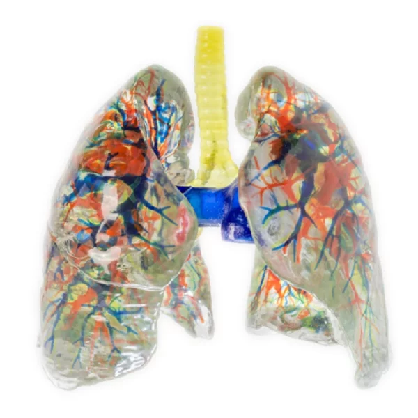
Digital Anatomical Architecture: Before touching any raw materials, medical engineers design the model using advanced 3D modeling software (often based on high-resolution CT or MRI scans of actual human anatomy). Every intricate detail is mapped out digitally—including the bronchial tree, alveoli clusters, pulmonary arteries, pulmonary veins, and the outer lung lobes.
Multi-Material 3D Printing & Color-Coded Precision: Once the digital blueprint is ready, high-precision industrial 3D printers get to work outputting individual components. Color choice is essential here for clear visual learning.
- Airways (Trachea & Bronchi): Printed in light/neutral shades (like off-white or light gray) to highlight the air pathway.
- Pulmonary Veins: Printed in vibrant blue to denote oxygen-depleted blood returning to the heart.
- Pulmonary Arteries: Printed in bright red to show oxygen-rich blood distribution.
- The Outer Shell / Body: Usually printed either in transparent resin so students can study the internal structures from every angle without taking it apart, or in a realistic soft pink to mirror actual living tissue.
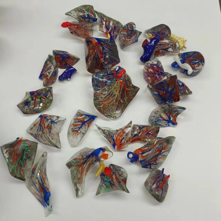
Assembly, Finishing, and Quality Control: For example, at DIGIHUMAN, The lung segment is segmented according to 8 segments of the left lung and 10 segments of the right lung to form a split and combined lung segment model. After printing, technicians carefully clean, polish, and assemble the individual printed parts into a complete, durable model. Once thoroughly tested for anatomical accuracy and durability, the finished models are packed up and shipped off to medical schools and laboratories worldwide.
Frequently Asked Questions
Q: How DIGIHUMAN ensures 3D lung model is anatomically precise?
A: DIGIHUMAN data is selected from a high-precision digital human dataset. Also, cadaveric specimens fixed by formalin were selected as a 3D printing basis for reference and comparison of the model.
Q: What materials are ideal for constructing a simple 3D lung model?
A: Common materials for children include a plastic bottle, balloons, straws, elastic bands, and play dough
Q: How DIGIHUMAN model?
A: The voxels of each anatomical structure surface were extracted from the original tomographic dataset to generate texture maps of the geometric model to ensure that the appearance of the geometric model of each anatomical structure has the same visual perception as that of the real anatomical specimen.
Conclusion
When talking about how to make 3D model of lung, whether it’s a simple plastic bottle experiment on a classroom desk or a hyper-realistic, color-coded 3D model in a medical lecture hall, these teaching tools bridge the gap between abstract science and hands-on understanding.
At DIGIHUMAN, we believe in the power of 3D models to enhance understanding and engagement in the field of anatomy. Explore our range of products and see how to make a 3d model of lungs and how we can support your educational endeavors today!

MEDICA 2026 in Germany: What Medical Training Technology Looks Like This Year
Medical schools are being asked to train more people than they can teach. The World Health Organization's first annual health...

What Is Augmented Reality Medical Training
Medical schools have a quiet math problem. A class of thirty students often shares a single cadaver, and the numbers...

Hierarchical Structure of Anatomy: Understanding the Human Body From Systems to Detailed Structures
The human body is a complex organization built through multiple levels of structure. From large body systems to tiny anatomical...