
Understanding the human brain requires a clear view of both individual structures and their connections. The brain contains complex regions that work together, making spatial understanding an important part of medical education. Traditional anatomy learning often relies on illustrations, models, and specimen observation. However, medical imaging has created new ways to explore internal structures.
The midsagittal section of the human brain CT scan provides a valuable perspective by showing the central plane of the brain and helping learners understand the relationship between different anatomical regions. With digital visualization developing in medical education, we are exploring how imaging data can support more practical and interactive anatomy learning.
Why the Midsagittal View Matters in Brain Anatomy Study
The midsagittal section shows the brain from a medial perspective by dividing it along the central line. This view allows learners to observe important structures that are difficult to understand from external views alone. Areas such as the corpus callosum, brainstem, cerebellum, and ventricular system can be studied in relation to their surrounding structures.
For medical students, understanding these relationships is an important step before moving into more advanced clinical subjects. A clear anatomical view helps learners connect structure with location and develop a stronger foundation for interpreting medical images.
CT and MRI technologies provide detailed image sequences that represent different layers of the human body. Instead of viewing anatomy as a single picture, students can observe changes between sections and understand how structures continue through different levels.
The use of the midsagittal section of the human brain CT scan in education helps demonstrate how imaging data can support anatomical knowledge. It allows learners to compare visual information from scans with three-dimensional concepts of brain organization.
Connecting Medical Imaging With Anatomical Understanding
Medical imaging is not only used in clinical environments but also plays an important role in healthcare education. Learning how to interpret images requires knowledge of anatomy, spatial relationships, and structural differences.
In traditional classrooms, students may need to imagine three-dimensional structures from two-dimensional materials. This process can be challenging, especially when studying organs with complicated internal arrangements like the brain.
Digital visualization provides another learning approach. By combining imaging data with interactive models, students can view structures from different angles and better understand how separate regions are connected.
At DIGIHUMAN, we focus on developing digital anatomy solutions based on real human body data reconstruction. Our goal is to support medical education by creating learning tools that connect anatomical knowledge with interactive exploration.
How Three-Dimensional Visualization Supports Brain Education
Three-dimensional models provide a different perspective compared with traditional images. Learners can rotate structures, adjust views, and observe relationships between different anatomical areas. These functions help explain complex topics during medical training.
In brain anatomy courses, interactive visualization can support both classroom teaching and independent study. Educators can highlight specific structures while explaining their positions and functions. Students can also review selected areas repeatedly to strengthen their understanding.
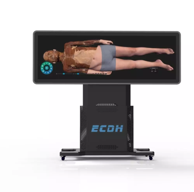
The HD Digihuman Virtual Anatomage Table applies real human body data reconstruction to simulate anatomy learning processes. The system displays human structures in 3D and allows users to zoom in, zoom out, and rotate models from different angles.
The platform also includes CT and MRI image sequences. Each case can contain basic information, imaging findings, and imaging diagnosis details. These resources help create a connection between medical imaging knowledge and anatomical observation.
Creating Flexible Teaching Experiences With Digital Anatomy Tools
Different medical institutions have different teaching requirements. Universities, training centers, and healthcare education organizations may need flexible tools for lectures, demonstrations, and student practice.
Modern digital anatomy systems provide functions that support customized teaching methods. Anatomical structures can be annotated, hidden, made transparent, separately displayed, locked, or selected according to learning needs. These options allow educators to focus on specific topics during lessons.
The HD Digihuman Virtual Anatomage Table includes an 88-inch display with a resolution of 3840×1080 and infrared touch technology. The display can be lifted vertically and reversed 90 degrees, allowing adjustment for different teaching environments.
Our system also supports user interaction management, marker creation, screenshots and screen recording, and correlated medical image resources. These functions provide additional support for organized anatomy education.
Exploring the Future of Brain Anatomy Visualization
The development of medical education is closely connected with advances in imaging and digital technology. Future anatomy learning will continue to focus on helping students understand structures through clearer, more interactive methods.
The combination of CT data, MRI information, and three-dimensional reconstruction provides a more complete way to study the human brain. The midsagittal section of the human brain CT scan is one example of how medical images can become educational resources rather than only diagnostic references.
We believe digital anatomy tools can support medical institutions that want to improve visualization-based teaching methods. Through solutions such as the HD Digihuman Virtual Anatomage Table, DIGIHUMAN continues to develop resources for anatomy education with real human body data and interactive technology.
By connecting imaging information with three-dimensional learning, we aim to help educators create clearer anatomy experiences. The development of digital visualization provides new opportunities for understanding complex structures, and the midsagittal section of the human brain CT scan continues to serve as an important reference for studying brain anatomy. We invite educators to explore how midsagittal brain CT views can be integrated into their own anatomy curricula with DIGIHUMAN.

Cadaver vs. Virtual Anatomy: How PA Students Learn the Human Body
Two routes now run through every physician assistant anatomy course. One starts with a donor on a steel table, the...

How Are Human and Rat Anatomy Similar?
When we study anatomy, one of the most interesting questions we encounter is how are human and rat anatomy similar?...

What Is the Largest Organ in the Rat’s Anatomy?
When we study animal anatomy, one of the simplest questions can lead to a surprisingly interesting discussion: what is the...