
Medical education has gradually moved from traditional illustrations and physical specimens toward digital learning resources. With the development of medical imaging technology, CT data has become an important reference for understanding the internal structures of the human body. A well-organized human pelvis CT scans archive can provide valuable learning materials for anatomy education, allowing students and educators to study complex structures through detailed sectional images and digital reconstruction.
The pelvis contains multiple bones, joints, muscles, and related anatomical structures that require careful observation during learning. Through digital image resources, learners can explore these structures from different perspectives and build a clearer connection between two-dimensional medical images and three-dimensional anatomy. At DIGIHUMAN, we focus on how digital technologies can support more interactive and practical approaches to medical education.
Understanding the Role of Pelvic CT Data in Anatomy Learning
CT imaging provides sectional views of the human body by capturing detailed internal structures. For anatomy students, these images help explain the relationship between bones, organs, and surrounding tissues. Compared with simple diagrams, medical image resources allow learners to observe real anatomical details and understand spatial connections more effectively.
The pelvis is an important area in anatomy education because it includes structures related to movement, support, and body balance. Studying pelvic CT data helps students recognize differences between individual anatomical layers and understand how various structures are arranged within a limited space.
A digital image collection can also support repeated learning. Students can review specific sections, compare different views, and strengthen their understanding through continuous observation. This learning method is useful for medical schools and training institutions that need reliable educational materials.
Digital Medical Image Resources and Modern Teaching Methods
The value of medical image resources is not only in storing images but also in creating a better learning process. Digital archives allow educators to organize imaging data according to teaching needs, making it easier to introduce specific anatomical topics during classes.
A human pelvis CT scans archive can include sectional images that help students connect CT visualization with actual anatomical structures. When these resources are combined with three-dimensional models, learners can move from image recognition to deeper structural understanding.
Modern anatomy education increasingly focuses on interaction. Instead of only viewing static materials, students can explore structures through digital platforms and participate in a more active learning process. This approach can help reduce the difficulty of understanding complicated anatomical relationships.
From Two-Dimensional Images to Three-Dimensional Anatomy Exploration
Traditional CT images are mainly presented as sectional slices, which require learners to imagine the complete structure from multiple views. Digital reconstruction technology changes this process by combining different image layers into a three-dimensional model.
Through three-dimensional visualization, students can observe anatomical structures from multiple angles. They can better understand the connection between different layers and improve their ability to interpret medical images. This transition from 2D observation to 3D exploration represents an important development in medical education.
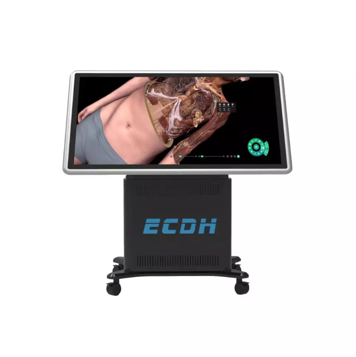
Based on real human body data reconstruction, our HD Digital Human Virtual Anatomy System Mini combines traditional anatomy teaching with digital technology. The system allows users to explore virtual anatomy operations while supporting different teaching environments through an adjustable screen that can be lifted and tilted.
Building Interactive Digital Anatomy Learning Environments
Digital anatomy systems provide more than visual models. They combine different learning resources, including three-dimensional structures, sectional images, CT/MRI images, teaching videos, and exercises. This combination creates a more complete learning environment for students and instructors.
Our HD Digital Human Virtual Anatomy System Mini includes the ECDH Anatomy System, which contains more than 3000 3D structures, more than 3000 sectional images, more than 1700 CT/MRI images, over 100 teaching videos, and more than 1800 exercises. These resources support teachers in organizing anatomy lessons and allow students to study structures in an interactive way.
The system also supports professional anatomy teaching software through a touch pad. Students can observe stereo human anatomy structures with 3D glasses and explore the virtual environment during learning activities. This type of technology provides another option for institutions looking to integrate digital resources into anatomy education.
The Future Value of Digital Medical Image Resources
As medical education continues to develop, digital image resources will play an important role in supporting anatomy teaching and professional training. The combination of real medical data, three-dimensional reconstruction, and interactive learning tools creates new possibilities for understanding human structures.
A carefully developed human pelvis CT scans archive represents more than a collection of medical images. It provides a foundation for exploring anatomy from different viewpoints and helps connect imaging knowledge with practical learning. Digital resources can support educators in creating more flexible teaching methods while giving students additional ways to understand complex structures.
Through our work at DIGIHUMAN, we continue exploring how digital anatomy technology can support medical education. Solutions such as the HD Digital Human Virtual Anatomy System Mini are designed to connect real human data with interactive learning experiences, helping educational institutions build modern anatomy teaching environments.

MEDICA 2026 in Germany: What Medical Training Technology Looks Like This Year
Medical schools are being asked to train more people than they can teach. The World Health Organization's first annual health...

What Is Augmented Reality Medical Training
Medical schools have a quiet math problem. A class of thirty students often shares a single cadaver, and the numbers...

Hierarchical Structure of Anatomy: Understanding the Human Body From Systems to Detailed Structures
The human body is a complex organization built through multiple levels of structure. From large body systems to tiny anatomical...