
Medical anatomy education requires students to understand not only the names of structures but also their positions, connections, and spatial relationships inside the human body. Traditional learning methods, including textbooks and physical specimens, provide important foundations, but some complex organs remain difficult to understand through two-dimensional materials alone.
With the development of digital visualization technology, educators now have more options to explain anatomy in a clearer way. In our experience, human male CT scan liver data offers valuable support for helping learners connect medical imaging information with three-dimensional anatomical knowledge.
Why CT Data Has Value in Liver Anatomy Education
The liver is one of the important organs studied in medical anatomy courses. Its structure includes different regions, vessels, and surrounding tissues that require careful observation. Students often need to understand how these parts are arranged rather than simply memorize individual names.
CT imaging provides sectional views of internal structures, allowing learners to observe the body layer by layer. These images contain detailed information that can support anatomy teaching when combined with suitable educational methods. By studying human male CT scan liver examples, students can gradually understand how cross-sectional images represent actual anatomical positions.
However, interpreting CT images can be challenging for students who are still developing spatial thinking skills. A series of separate images may not immediately show the complete relationship between different structures. This is where digital reconstruction can provide additional support.
Digital anatomy technology transforms medical data into interactive three-dimensional models. Students can view structures from multiple directions and compare different anatomical layers. This approach helps connect image-based learning with a more complete understanding of human anatomy.
Combining Medical Imaging With Three-Dimensional Learning
Modern medical education is increasingly focused on creating learning environments that encourage observation and exploration. Anatomy is not only about identifying structures but also about understanding how these structures exist together within the body.
Three-dimensional visualization allows students to explore organs in a more flexible way. They can observe external forms, examine internal relationships, and review specific areas based on their learning needs. This process supports a deeper understanding compared with only viewing static diagrams.
For educators, digital anatomy tools provide additional teaching methods. Teachers can use interactive models during lectures, explain complicated structures step by step, and create demonstrations that match different learning objectives.
The combination of medical imaging and digital reconstruction also helps students build connections between classroom knowledge and future clinical learning. When learners understand how imaging information reflects human anatomy, they can develop stronger foundations for interpreting medical images in later studies.
Improving Learning Efficiency Through Interactive Anatomy Platforms
Digital anatomy platforms are designed to support active learning. Instead of only receiving information from instructors, students can interact with anatomical models and explore structures independently. This creates more opportunities for observation and discussion during education.
Interactive learning is especially useful for organs with complex spatial relationships. The liver, for example, requires an understanding of different sections and surrounding structures. Using human male CT scan liver resources in a digital environment allows students to compare sectional information with reconstructed anatomical views.
In medical institutions, these tools can support different teaching scenarios, including classroom instruction, laboratory demonstrations, and professional training programs. They provide educators with flexible resources that can be adjusted according to course requirements.
At DIGIHUMAN, we focus on developing digital anatomy solutions that connect medical visualization resources with interactive learning technologies. Our goal is to support educational organizations that need reliable tools for anatomy teaching and help create more practical learning experiences.
Exploring the Development of Digital Anatomy Technology
The growth of digital anatomy reflects changes in medical education methods. As technology continues to improve, more institutions are exploring ways to combine medical data, three-dimensional models, and interactive systems.
A digital anatomy system needs accurate data and suitable visualization methods to present structures clearly. High-quality reconstruction plays an important role because realistic models help learners recognize anatomical details and understand relationships between different areas.
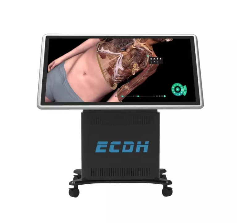
The Digihuman Virtual Anatomage Table integrates medical visualization resources and human-computer interaction technologies. It combines gross anatomy with the Digihuman Anatomy System to create a virtual training platform. Based on ultra-high-precision human tomographic data with 0.1 mm reconstruction accuracy and advanced 3D reconstruction technology, the system provides realistic anatomical structures and teaching functions.
This type of platform can help universities, medical schools, and training organizations explore new approaches to anatomy education. It supports interactive learning while maintaining a connection with traditional anatomical knowledge.
Creating New Possibilities for Future Medical Education
Digital anatomy does not replace traditional education methods. Instead, it provides additional resources that help teachers explain complex concepts and allow students to explore structures in different ways.
As medical education continues to develop, the connection between imaging technology and anatomy learning will become increasingly important.
Through continuous development of digital visualization solutions, DIGIHUMAN supports institutions in building modern anatomy learning environments. By applying resources such as human male CT scan liver within interactive platforms, we help educators provide clearer explanations of anatomical structures and support students in developing stronger three-dimensional understanding.

MEDICA 2026 in Germany: What Medical Training Technology Looks Like This Year
Medical schools are being asked to train more people than they can teach. The World Health Organization's first annual health...

What Is Augmented Reality Medical Training
Medical schools have a quiet math problem. A class of thirty students often shares a single cadaver, and the numbers...

Hierarchical Structure of Anatomy: Understanding the Human Body From Systems to Detailed Structures
The human body is a complex organization built through multiple levels of structure. From large body systems to tiny anatomical...