
The pelvic region is one of the more complex areas of human anatomy because it contains multiple structures that work together within a limited space. Bones, muscles, vessels, nerves, and organs are closely connected, making it important for medical professionals and educators to understand their spatial relationships clearly.
With the development of digital visualization technology, anatomy education has gained new methods for presenting complex structures. When studying human pelvis CT scans, educators can combine imaging knowledge with three-dimensional anatomical models to create a more complete learning experience.
Understanding the Complexity of Pelvic Structures
The pelvis plays an important role in supporting the body and connecting different anatomical systems. Its structure includes the pelvic bones, surrounding soft tissues, blood vessels, and internal organs. Because these components overlap in traditional images, understanding their positions can require strong spatial imagination.
In medical education, teachers often need to explain how different structures relate to each other rather than only showing individual parts. Three-dimensional visualization provides a clearer way to demonstrate depth, location, and connections between anatomical regions.
CT imaging provides detailed information about internal structures, but medical learners still need to translate two-dimensional images into a three-dimensional understanding. This process requires practice and professional guidance. Digital anatomy resources can help bridge the gap between image interpretation and structural recognition.
Connecting Medical Imaging With 3D Anatomy Learning
Medical imaging has become an important part of modern healthcare education. CT scans allow professionals to observe internal structures without traditional physical exploration. However, interpreting cross-sectional images requires knowledge of anatomy and experience with spatial reconstruction.
For example, human pelvis CT scans can show different tissue layers and structural details through multiple sections. By combining these images with interactive 3D models, learners can better understand how each section corresponds to the complete anatomical structure.
This connection is valuable for medical schools, training centers, and professional education programs. It allows educators to explain complicated anatomical relationships through visual demonstrations instead of relying only on textbook illustrations or static images.
Digital Tools Support More Detailed Anatomy Presentation
As medical education continues to develop, digital anatomy platforms provide additional resources for teachers and learners. These tools allow users to explore structures from different angles and observe relationships that may be difficult to display through traditional teaching materials.
At this stage, we have developed solutions that focus on presenting anatomy in a structured and visual way. DIGIHUMAN provides digital anatomy resources that support systematic anatomy, regional anatomy, and sectional anatomy learning. These approaches help educators organize teaching content according to different educational requirements.
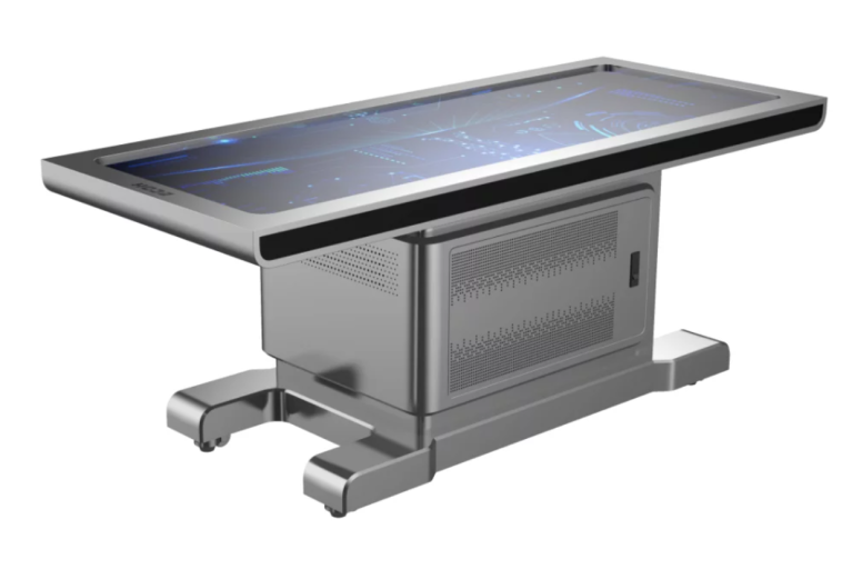
The Digihuman Virtual Anatomage Table includes more than 6000 3D anatomical structures. It also provides real dissection videos and anatomy animations, allowing users to observe anatomical processes through different forms of content. In addition, test question resources are available to support knowledge review and consolidation.
Improving the Interpretation of Complex Anatomical Information
One challenge in anatomy education is helping learners move from memorizing names to understanding functional relationships. A structure-based learning approach allows users to explore anatomy through location, connection, and interaction.
For spinal anatomy, this method is especially useful because many important structures are located close together. Digital models can help show how bones, muscles, and soft tissues are arranged, making explanations more direct during classroom demonstrations.
Educators can also adjust teaching methods based on different learning stages. Beginners may focus on basic structures, while advanced learners can explore more detailed anatomical relationships. This flexibility supports different teaching environments and professional training needs.
The Role of Visualization in Future Medical Education
The use of digital visualization does not replace traditional anatomy education. Instead, it provides another way to present information and support deeper understanding. Combining physical learning resources, medical imaging, and interactive models can create a more comprehensive educational process.
As technology continues to develop, medical institutions are paying more attention to how digital resources can improve teaching efficiency and content presentation. Clear visualization methods may become an important part of anatomy education, especially for areas where structures are highly connected.
Through continuous exploration of digital anatomy applications, DIGIHUMAN focuses on providing educational tools that help institutions present anatomical knowledge in a more organized and interactive format. These solutions can support teachers in explaining complex topics and allow learners to explore structures with greater clarity.
Exploring New Possibilities for Anatomy Education
Understanding the human body requires both detailed information and effective ways to present that information. From medical imaging interpretation to three-dimensional exploration, different learning methods can work together to improve anatomical knowledge.
The development of digital anatomy platforms provides new opportunities for medical education organizations. By integrating visualization, educational resources, and structured learning methods, institutions can create more flexible teaching environments. In this process, human pelvis CT scans can become easier to understand when combined with three-dimensional anatomical references, helping connect imaging knowledge with practical anatomy learning.
With its focus on digital anatomy technology, DIGIHUMAN continues to provide resources for medical education and professional training. Through tools such as the Digihuman Virtual Anatomage Table, educators can introduce detailed anatomical structures, animations, and learning materials to support different teaching scenarios.

MEDICA 2026 in Germany: What Medical Training Technology Looks Like This Year
Medical schools are being asked to train more people than they can teach. The World Health Organization's first annual health...

What Is Augmented Reality Medical Training
Medical schools have a quiet math problem. A class of thirty students often shares a single cadaver, and the numbers...

Hierarchical Structure of Anatomy: Understanding the Human Body From Systems to Detailed Structures
The human body is a complex organization built through multiple levels of structure. From large body systems to tiny anatomical...