
Understanding animal anatomy is essential for veterinary medicine, animal science, and responsible pet care. Among the many areas of veterinary anatomy, female dog anatomy deserves particular attention because the reproductive system has a distinctive structure and plays an important role in reproduction, pregnancy, and clinical health.
At DIGIHUMAN, we work with digital anatomy and medical visualization technologies. Although our primary focus is human anatomy education, we recognize that the principles of anatomical visualization are valuable across biological sciences. In this article, we will explore the basic structures of female dog anatomy, with particular attention to the reproductive system and its relationship with the rest of the body.
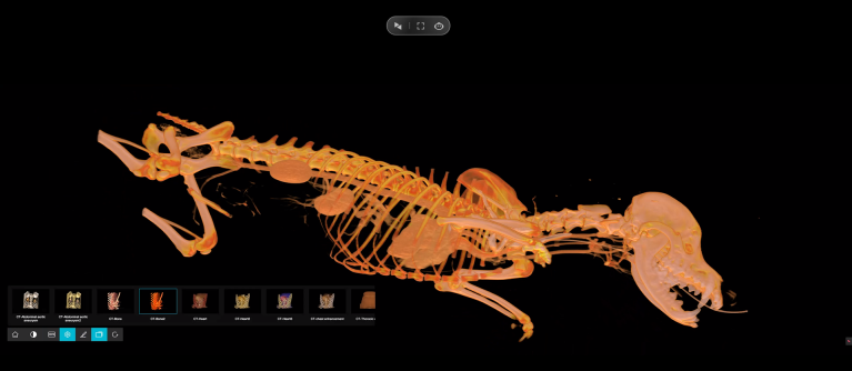
What Makes Female Dog Anatomy Different?
The anatomy of a female dog includes the same broad body systems found in other mammals, including the skeletal, muscular, digestive, respiratory, cardiovascular, nervous, and reproductive systems. However, each species has anatomical characteristics that reflect its biology and reproductive behavior.
When studying female dog anatomy, the reproductive system is often one of the most important areas to understand. The female genital tract includes the ovaries, oviducts, uterus, cervix, vagina, and vulva. The mammary glands are also associated with the reproductive system.
These structures do not function independently. Instead, they form a connected system that supports the production of eggs, fertilization, pregnancy, and birth.
The Ovaries and Their Role in Reproduction
The ovaries are important organs in female dog anatomy because they produce ova, or eggs, and secrete reproductive hormones.
The female reproductive cycle is influenced by hormones including estrogen and progesterone. These hormones are closely associated with changes in reproductive function throughout the cycle. Veterinary literature describes the reproductive system as an integrated structure in which the ovaries, reproductive tract, and hormones work together.
The ovaries are connected to the oviducts, also known as Fallopian tubes. After ovulation, an egg can travel through an oviduct toward the uterus. The oviduct therefore serves as an important connection between the ovary and the uterine region.
The Uterus and Its Role in Pregnancy
Another central structure in female dog anatomy is the uterus. The uterus provides the environment in which embryos can develop into fetuses during pregnancy.
The canine uterus has two uterine horns that connect toward the central uterine body. This structure is particularly relevant to canine reproduction because developing embryos are located within the uterine environment before birth.
At the end of the uterus is the cervix. The cervix forms a connection between the uterus and vagina and serves as an important anatomical barrier between the reproductive tract and the outside environment.
Understanding these relationships is particularly useful for veterinary students because reproductive anatomy provides the foundation for studying pregnancy, parturition, reproductive disorders, and related clinical procedures.
Vagina and Vulva: Understanding Internal and External Structures
The vagina is a muscular tube that extends from the cervix toward the external reproductive opening. The vulva forms the external part of the female genital tract.
This distinction is important when studying female dog anatomy because internal and external structures have different anatomical positions and functions.
For veterinary learners, understanding their spatial relationship can also make it easier to interpret clinical examinations and diagnostic images. Anatomy is not simply a list of individual organs; it is fundamentally about understanding where structures are located and how they connect with one another.
Why 3D Visualization Can Help With Anatomy Education
One challenge in anatomy education is understanding three-dimensional relationships from flat diagrams. A textbook illustration may show an organ clearly, but students still need to mentally translate that image into a three-dimensional structure.
This is where digital visualization can provide an additional learning approach. Interactive three-dimensional models allow learners to rotate structures, change viewing angles, and examine relationships between different anatomical components.
At DIGIHUMAN, we apply this visualization principle to human medical education. Our digital anatomy technologies are designed to help learners explore anatomical structures in a more interactive way.
Our Virtual Anatomy Table organizes anatomy into systematic, regional, and sectional approaches and includes more than 6,000 three-dimensional anatomical structures, along with dissection videos, anatomy animations, and learning resources.
Although these resources are designed around human anatomy rather than female dog anatomy, the educational principle is similar: interactive visualization can help students develop a clearer understanding of spatial relationships within the body.
Why Understanding Anatomy Matters
Learning female dog anatomy is more than memorizing the names of reproductive organs. It involves understanding how structures are positioned, how they connect, and how they work together as a biological system.
For veterinary professionals and students, this knowledge can support the study of reproduction, diagnostic imaging, surgery, pathology, and animal health. For researchers and educators, accurate anatomical visualization can also make complicated biological concepts easier to communicate.
At DIGIHUMAN, we continue to develop digital anatomy solutions that make anatomical education more visual and interactive. Through our virtual anatomy tables and other digital teaching systems, we aim to support medical and scientific education by helping learners move from simply memorizing structures to understanding their three-dimensional relationships.

Cadaver vs. Virtual Anatomy: How PA Students Learn the Human Body
Two routes now run through every physician assistant anatomy course. One starts with a donor on a steel table, the...

How Are Human and Rat Anatomy Similar?
When we study anatomy, one of the most interesting questions we encounter is how are human and rat anatomy similar?...

What Is the Largest Organ in the Rat’s Anatomy?
When we study animal anatomy, one of the simplest questions can lead to a surprisingly interesting discussion: what is the...