
When we look at a dog running, jumping, turning, or simply standing, it is easy to focus on the muscles and movement we can see. However, the foundation of these actions lies beneath the surface. Understanding dog skeleton anatomy helps us appreciate how a canine body supports weight, protects vital organs, and produces coordinated movement.
At DIGIHUMAN, we believe that anatomy becomes easier to understand when complex structures can be explored visually and systematically. Although our core digital anatomy solutions are designed primarily for medical education, the principles of three-dimensional anatomical visualization also provide an interesting way to think about skeletal structures and spatial relationships.
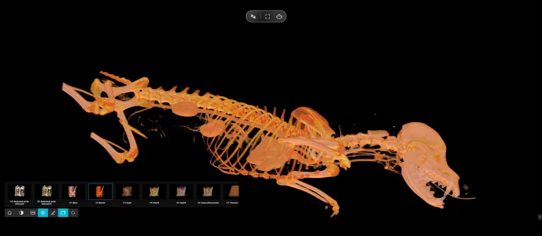
What Does Dog Skeleton Anatomy Include?
The canine skeleton is a structural framework made up of bones that support the body and protect internal organs. Bones also contain bone marrow and serve as important reservoirs for minerals such as calcium and phosphorus. As with other mammals, canine bone tissue is continuously remodeled, with older tissue replaced by new tissue.
For a basic study of dog skeleton anatomy, we can divide the skeleton into several major regions: the skull, vertebral column, ribs and sternum, forelimbs, and hindlimbs.
The skull protects the brain and forms the structural foundation of the face and jaws. Behind it, the vertebral column extends from the neck toward the tail. The spinal column is composed of successive vertebral regions—cervical, thoracic, lumbar, sacral, and caudal—which work together to provide support while allowing controlled flexibility.
The ribs and associated structures help protect organs within the chest, while the bones of the limbs form the framework needed for locomotion.
How Do Bones Help Dogs Move?
Bones do not work independently. They connect at joints, while muscles, tendons, ligaments, and cartilage contribute to movement and stability. The shape and structure of a joint influence the direction and degree of movement it can provide. For example, some joints primarily permit bending and straightening, while others allow greater rotational movement.
This relationship is particularly important when studying dog skeleton anatomy because canine locomotion depends on coordinated interaction between multiple anatomical structures.
The forelimbs and hindlimbs have different mechanical roles, yet they function together during walking and running. The spine also contributes to overall movement by providing a flexible but supportive connection between the head, trunk, and limbs.
This is why studying individual bones in isolation can provide only part of the picture. To understand canine movement properly, learners need to consider how bones connect with joints and how these structures relate to surrounding muscles and connective tissues.
Why Is Dog Skeleton Anatomy Important to Study?
A clear understanding of skeletal anatomy is useful in veterinary education, animal science, rehabilitation, and clinical practice. It establishes the anatomical foundation for understanding conditions involving bones and joints.
According to the Merck Veterinary Manual, musculoskeletal disorders can affect a dog’s movement, and problems may arise from trauma, developmental abnormalities, nutritional factors, inherited conditions, infections, or tumors.
For students, visualizing the relationship between bones and joints can therefore make later topics easier to understand. Instead of memorizing isolated anatomical names, learners can build a mental three-dimensional model of how structures fit together.
This approach is also valuable when studying diagnostic images. Understanding the normal position and relationship of bones provides an essential reference point when interpreting abnormalities.
From Traditional Anatomy to Digital Visualization
Anatomy education has traditionally relied on textbooks, illustrations, physical specimens, radiographic images, and anatomical models. Digital visualization adds another layer by allowing learners to manipulate three-dimensional structures and examine them from different perspectives.
We see this as particularly valuable for teaching spatial relationships. A two-dimensional diagram may show the position of a bone clearly, but an interactive three-dimensional model can allow a learner to rotate, enlarge, isolate, or compare structures.
At DIGIHUMAN, our virtual anatomy approach follows this principle. Our Virtual Anatomy Table categorizes anatomy into systematic, regional, and sectional views and provides more than 6,000 three-dimensional anatomical structures, alongside dissection videos, anatomy animations, and question resources.
The broader educational value is not simply the technology itself. It is the ability to move between different views and levels of anatomical detail, helping learners connect structure with function.
What Can We Learn From a 3D Approach?
The study of dog skeleton anatomy highlights an important principle in anatomy education: structures are easier to understand when their relationships are visible. Bones provide support and protection, joints enable controlled movement, and muscles transmit force through the skeletal framework. Understanding how these structures work together helps learners connect anatomical structure with function.
Interactive 3D visualization allows learners to examine anatomical structures from different perspectives, explore their spatial relationships, and better understand how individual components form an interconnected system. This approach can make complex anatomical concepts easier to understand and provide a useful foundation for further study in anatomy and related fields.
Enhancing Anatomy Education with DIGIHUMAN
At DIGIHUMAN, we develop digital anatomy technologies to help educators, students, medical professionals, and researchers explore anatomical structures through interactive visualization. Our product portfolio includes virtual anatomy tables, anatomy teaching systems, VR and AR anatomy systems, 3D anatomical models, and digital morphology solutions.
Our Virtual Anatomy Table features more than 6,000 three-dimensional anatomical structures, alongside dissection videos, anatomy animations, and assessment resources. By enabling users to explore anatomy through systematic, regional, and sectional views, our solutions support more interactive learning experiences and help educational institutions make complex anatomical knowledge more accessible.

Cadaver vs. Virtual Anatomy: How PA Students Learn the Human Body
Two routes now run through every physician assistant anatomy course. One starts with a donor on a steel table, the...

How Are Human and Rat Anatomy Similar?
When we study anatomy, one of the most interesting questions we encounter is how are human and rat anatomy similar?...

What Is the Largest Organ in the Rat’s Anatomy?
When we study animal anatomy, one of the simplest questions can lead to a surprisingly interesting discussion: what is the...