
When we watch a dog run, jump, climb stairs, or simply stand, it is easy to see the movement but not the complicated anatomical system making it possible. Dog leg anatomy involves bones, joints, muscles, tendons, and ligaments working together to provide support, stability, and movement. Understanding how these structures interact can help students, educators, veterinary professionals, and curious pet owners better appreciate canine locomotion.
At DIGIHUMAN, we believe that anatomy becomes easier to understand when complex structures can be explored visually and systematically. Although our current virtual anatomy products focus on human anatomical data, the principles of digital anatomy education provide an interesting way to think about how three-dimensional anatomical structures can be studied.
What Are the Main Parts of Dog Leg Anatomy?
A dog’s limbs can be divided into forelimbs and hindlimbs, and their structures have different anatomical relationships and functions. The hindlimb is particularly important for propulsion. According to the University of Calgary’s veterinary anatomy resources, the canine pelvic limb connects to the axial skeleton through the pelvis, while major muscle groups around the limb contribute to flexion and extension.
When studying dog leg anatomy, it is also important to understand that the word “leg” can have a narrower anatomical meaning than it does in everyday conversation. In veterinary anatomy, the leg may refer specifically to the region between the stifle and hock, corresponding mainly to the tibia and fibula. The broader hindlimb includes the pelvis, femur, lower leg, and paw.
This distinction matters because different bones and joints perform different mechanical roles.
How Do the Bones Support a Dog’s Movement?
The hindlimb contains several major skeletal structures. The pelvic girdle includes the ilium, ischium, and pubis. Moving downward, the femur forms the thigh, while the tibia and fibula make up the lower leg. The distal limb includes the tarsal bones, metatarsals, and phalanges.
These bones provide structural support while also forming joints that allow controlled movement. Merck Veterinary Manual explains that bones provide rigid structural support, while joints determine the direction and degree of movement. Cartilage at joint surfaces helps reduce friction as bones move against one another.
The stifle, often compared with the human knee, is especially important in canine locomotion. Together with the hip and hock, it allows the hindlimb to move through coordinated flexion and extension.
What Role Do Muscles, Tendons, and Ligaments Play?
Bones cannot move by themselves. Muscles generate force, while tendons transfer that force from muscles to bones. Ligaments connect bones to other bones and help stabilize joints. This relationship is fundamental to dog leg anatomy and explains why healthy movement depends on more than the skeletal structure alone.
Several muscle groups contribute to hindlimb movement. The quadriceps are located toward the front of the thigh and contribute to extension of the stifle. The hamstring group lies toward the back of the thigh and contributes to flexion and propulsion. Veterinary anatomy references also describe gluteal muscles as important contributors to hip movement.
Lower-leg muscles influence the hock and digits. For example, the gastrocnemius extends the hock, while other muscles control movements of the paw and toes. The Achilles tendon is a strong structure running toward the hock and incorporates tendons from several muscles.
Why Is Three-Dimensional Anatomy Useful for Learning?
One challenge in anatomy education is understanding the relationship between structures located at different depths. A flat illustration can identify a muscle or bone, but learners may find it harder to visualize how that structure relates spatially to neighboring tissues.
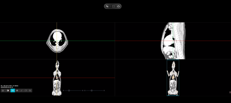
This is one reason we at DIGIHUMAN emphasize interactive digital anatomy. Our virtual anatomy table allows anatomical structures to be displayed in three dimensions and manipulated through functions such as enlargement, reduction, rotation, hiding, transparency, separate display, and multi-selection. The system also integrates CT and MRI image sequences for anatomy-related teaching and case exploration.
Our HD DIGIHUMAN Virtual Anatomy Table comes with a large infrared touchscreen that can be electrically adjusted and rotated. Equipped with high-performance hardware and certified with CE and FCC, it supports interactive 3D anatomical viewing.
It is important to clarify that this product is built around reconstructed human anatomical data, not a canine anatomy database. Therefore, we would not describe it as a dedicated dog anatomy teaching system. Instead, it demonstrates how interactive visualization can make complicated anatomical relationships easier to explore.
Bringing Anatomy Learning Into the Digital Age
Studying dog leg anatomy illustrates a broader principle: anatomy is not simply a list of bones and muscles. It is a dynamic system in which structure and function are closely connected. When a dog runs, muscles generate force, tendons transmit that force, joints guide movement, and bones provide mechanical support.
At DIGIHUMAN, we apply the same commitment to visual clarity and structured anatomical learning to human anatomy education. Our virtual anatomy solutions are designed around reconstructed anatomical data and interactive visualization, helping medical educators and students explore structures beyond the limitations of conventional two-dimensional materials.

Cadaver vs. Virtual Anatomy: How PA Students Learn the Human Body
Two routes now run through every physician assistant anatomy course. One starts with a donor on a steel table, the...

How Are Human and Rat Anatomy Similar?
When we study anatomy, one of the most interesting questions we encounter is how are human and rat anatomy similar?...

What Is the Largest Organ in the Rat’s Anatomy?
When we study animal anatomy, one of the simplest questions can lead to a surprisingly interesting discussion: what is the...