
The anatomy lab has long been an essential venue for cultivating medical students’ practical skills. However, it also faces limitations that are difficult to ignore.
For example, with the expansion of the educational scale, the limited cadaver sourcing, coupled with strict ethical reviews and donation procedures, makes it difficult to meet teaching demands. Additionally, long-term preservation requires expensive chemical reagents (such as formaldehyde) and constant-temperature facilities, resulting in high maintenance costs.
How can these problems be addressed? Virtual dissection tables emerged as an effective solution. Through high-precision 3D modeling and interactive software, students gain unlimited access to digitized specimens of human anatomical structures.
This blog will discuss more about virtual dissection tables, including benefits and applications. Read on to learn more!
What is a Virtual Dissection Table?
The virtual dissection table, also known as the digital anatomy table or 3D virtual anatomy table, is an advanced hardware-software integrated platform that utilizes 3D technology to create interactive, high-resolution anatomical models. These 3D models allow users to explore human anatomical structures in a dynamic and immersive environment.
A virtual dissection table operates as a seamlessly integrated system consisting of specialized hardware and custom 3D software:
- Hardware Architecture: The hardware is structured around a large, multi-touch interactive display mounted on a heavy-duty chassis with an embedded high-performance computing unit to render complex 3D volumetric models smoothly in real time. The touch display simulates the dimensions of an operating or dissection table to renders human anatomy at a 1:1 scale. It also supports intuitive gesture recognition—including single-finger dragging, two-finger zooming and rotating, and multi-finger slicing or isolating.
- Software Architecture: The software environment turns real medical imaging into an interactive digital lab. It is built from digitized, high-resolution cross-sectional scans of real human cadavers. The interactive programming allows users to cut, slice, layer, and reconstruct anatomical systems in real time across multiple planes.
The 3D virtual dissection table acts as an innovative tool that addresses the shortcomings of traditional anatomy teaching.
- Safe and Risk-Free
In traditional anatomy experiments, cadavers or organs need to be treated with formaldehyde and stored in specialized preservation equipment. Each classroom demonstration requires reopening the specimens. Repeated operations can easily cause wear, and improper handling by students may even damage the specimens.
However, the virtual dissection table eliminates exposure to harmful chemicals (such as formaldehyde) or biohazard risks associated with traditional dissection, ensuring the health of both teachers and students. Additionally, the digital models in the virtual dissection table cannot be damaged, allowing users to perform dissections repeatedly without worrying about specimen degradation.
- Efficient Teaching Outcomes
Digital anatomy tables are also crucial for enhancing overall teaching outcomes. Students can view anatomical structures from all angles, rotate them, and zoom in. They can also manipulate the 3D model to get a good grip on complicated concepts. Cross-sectional views and physiological human anatomy simulations further lead to spatial reasoning and knowledge retention.
What are the Applications of the Virtual Dissection Table?
The applications of virtual dissection tables are diverse, ranging from medical education to clinical training:
- Medical Education: It includes anatomy teaching and pathology learning, helping students understand systemic anatomy, regional anatomy, and sectional anatomy. It can also display various clinical cases, including basic information, chief complaints, imaging findings, and diagnoses, thereby promoting students’ application of theoretical knowledge to clinical practice.
- Clinical Training: Physicians can use 3D virtual dissection tables to explain different diseases and procedures to patients, improving communication. Moreover, the virtual dissection of the human bodyallows surgeons to rehearse important surgeries and improve their skills.
The advantages of DIGIHUMAN HD Virtual Anatomy Table
- Internationally Recognized Certifications
Safety, compliance, and product reliability are critical for medical and educational equipment. The Digihuman Virtual Anatomy Table has successfully obtained both CE Certification (European Conformity) and FCC Certification (Federal Communications Commission).
- Excellent Display Performance
Engineered to match the physical proportions of a real cadaver dissection bed, the table features an impressive 88-inch ultra-wide display. Its resolution is 3840×1080 panoramic clarity, and brightness is 700 cd / ㎡, ensuring crisp, clear visuals even under bright laboratory or lecture hall lighting. The contrast ratio is 1100:1 for deep blacks and vivid differentiation of tissue layers.
- Infrared multi-touch technology
Our virtual autopsy table supports smooth, multi-user gesture interactions (single-finger rotation, two-finger zooming, three-finger panning, five-finger operation panel positioning, and long-press cancel).
- Ergonomic Multi-Angle & Motorized Positioning
To adapt to diverse teaching environments—such as small group dissection labs, large lecture halls, or clinical demonstrations—this table is equipped with flexible physical adjustment controls. The table height can be raised or lowered to suit standing or seated users. The screen surface can be adjusted from a flat horizontal plane to tilted angles. Moreover, it supports switching between horizontal (0°) and vertical (90°) orientations.
- Workstation-Grade Hardware Specification
Rendering high-density volumetric datasets in real time without latency requires massive processing power. The built-in workstation is configured with high-end hardware, including Intel Core i7 (10th Generation or higher) processor, 64 GB DDR4 3200 MHz RAM memory, 2 TB NVMe SSD storage, NVIDIA GeForce RTX 3080 (10 GB VRAM or higher) graphics, and Windows 10 Professional operating system
- 6. R&D Excellence and Native Multilingual Support
The software architecture powering the table is the product of years of dedicated research by Digihuman’s expert R&D team, working in close collaboration with distinguished professors, medical researchers, and senior clinicians from leading institutions worldwide.
Our software is built upon tens of thousands of real medical data points gathered across global hospitals and research centers, featuring a native English software interface to support international medical curricula and global academic partnerships.erface to support international medical curricula and global academic partnerships.
- Ultra-High-Resolution Real Human Datasets
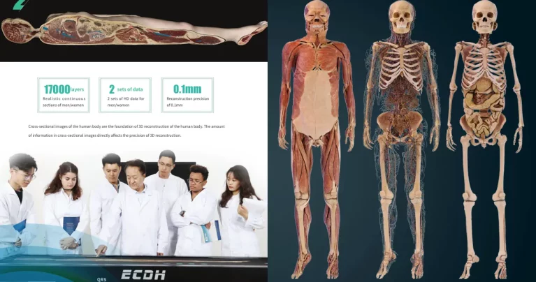
The Digihuman system sets a new standard for anatomical fidelity by utilizing ultra-high-resolution real human cadaver scan data rather than stylized or hand-drawn 3D graphics. The platform incorporates complete male and female datasets engineered for maximum scientific accuracy, with the male dataset comprising over 17,000 total cross-sectional layers at a section resolution of 13700 × 6340, and the female dataset featuring over 16,000 total cross-sectional layers at a section resolution of 12000 × 5700. Built with a single pixel scale of 26000× 16000× 3, both datasets achieve a sub-millimeter reconstruction precision of 0.1 mm, ensuring that fine structural elements such as micro-vasculature, peripheral nerve branches, and delicate fascial layers are preserved and rendered with complete anatomical realism.
- Interaction & Tools
The Digihuman system provides a powerful suite of intuitive dissection tools—including real-time rotation, smooth continuous panning and zooming, arbitrary multi-angle cutting, and interactive brush and dyeing functions—that make virtual cadaver dissection remarkably effortless, highly engaging, and entirely repeatable for both educators and students.
- Comprehensive Built-in Educational Resource Suite
Beyond basic 3D gross anatomy, the table integrates a vast digital teaching suite featuring over 3,000 detailed 3D anatomical structures, 3,000 tomographic slice images, 1,700 real clinical CT and MRI image series, 100 instructional videos, and more than 1,800 built-in practice exercises, offering an all-in-one ecosystem for lecture instruction, clinical visualization, and student assessment.

Where to buy virtual dissection tables?
If you are searching for reliable virtual dissection tables, look no further than the Digihuman virtual anatomy table. Digihuman was founded in 2002 and has been deeply engaged in the field of medical education products, particularly Digihuman anatomy systems and tables. We collaborate with experts from leading Chinese medical schools to develop a series of innovative products, including Virtual Anatomy Table, Anatomy System, 3D Printing Model, Morphology.
Our products are exported overseas, and have been affirmed by customers in more than 40 countries, serving medical education and training by providing an immersive and interactive learning experience for physicians, medical students, and researchers. Contact – Shandong Digihuman Technology Co., Inc with us today to enhance learning and research!

How Human Anatomy VR for Institutions Is Transforming the Modern VR Anatomy Teaching System
Human anatomy visualization has evolved from traditional methods to more digital and interactive approaches. Today, Human Anatomy VR for institutions provides...

Why Is Medical Simulation Important? From Cadaver Dissection to Virtual Anatomy Tables
Medical simulation has become an essential part of healthcare education, allowing learners to develop practical skills before treating real patients....

3D Printed Anatomical Model Explained: Concept, Applications, and Advantages
Advances in 3D printing and digital modeling are reshaping many fields. One of the most visible changes can be seen...