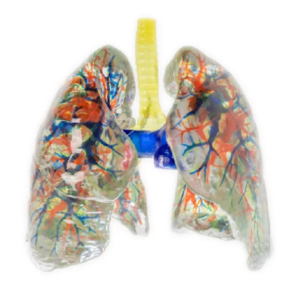
In the evolving landscape of medical education, precision and realism are crucial. At DIGIHUMAN, we are dedicated to providing high-quality educational tools that enhance understanding of human anatomy. Our lung anatomical model exemplifies this commitment, constructed with meticulous attention to detail and accuracy.
Comprehensive Data Sources
The development of our lung anatomical model begins with the selection of high-precision data sources. We utilize a robust digital human dataset that includes original tomographic data and refined segmentation data. This dataset captures the intricate details of lung anatomy, including various structures such as bones, muscles, blood vessels, nerves, and ligaments.
The voxel size of this dataset is 0.0384mm x 0.0384mm x 0.1mm, allowing for exceptional detail in the 3D model. Additionally, we incorporate cadaveric specimens that have been fixed with formalin. These specimens serve as a vital reference for comparison, ensuring that our lung anatomical model accurately reflects real human anatomy. By leveraging these diverse data sources, we can create a comprehensive and realistic representation of the lung’s complex structures.
Advanced Modeling Techniques
Once the data sources are established, we apply advanced modeling techniques to create our lung anatomical model. The voxels of each anatomical structure’s surface are meticulously extracted from the original tomographic dataset. This process allows us to generate detailed texture maps for the geometric models, ensuring that the appearance of our lung anatomical model closely resembles that of real anatomical specimens.
Our full-color, multi-material 3D printing technology further enhances the realism of the lung anatomical model. We use environmentally friendly resin materials to produce a 1:1 high simulation of the anatomical structures. This attention to detail not only provides a visual representation but also allows users to engage with the model tactilely, enhancing the learning experience.
Enhancing Medical Education
The lung anatomical model we offer is more than just a visual aid; it serves as a powerful educational tool. By integrating high-precision data and advanced modeling techniques, we provide students and educators with a realistic resource for studying lung anatomy. This model facilitates a deeper understanding of anatomical relationships and functions, making it invaluable for medical students, healthcare professionals, and educators.
In conclusion, the development of our lung anatomical model at DIGIHUMAN represents a significant advancement in anatomical education. By utilizing high-quality data sources and sophisticated modeling techniques, we create accurate and detailed representations of human anatomy. We encourage educational institutions to incorporate our lung anatomical model into their curricula, enriching the learning experience and preparing future healthcare professionals for success.

Revolutionizing Medical Education: Understanding the True Value and Human Anatomy VR Price
As pioneers in advanced medical virtualization, we at DIGIHUMAN have witnessed a fundamental shift in how anatomical knowledge is imparted...

Revolutionizing Medical Education: Why Human Anatomy VR for Institutions is Essential
As medical education evolves, the need for innovative and highly accurate teaching tools has never been greater. For years, educators...
Transforming Medical Education: The Power of Anatomy Physiology VR
In the specialized field of medical education, the foundational study of human anatomy and physiology has always been paramount. However,...