Advances in 3D printing and digital modeling are reshaping many fields. One of the most visible changes can be seen in medical education. Traditional anatomy education mainly relies on cadaver specimens, but these resources are limited. Securing and preserving them is also expensive.
3D printed anatomical models offer a practical solution to this problem, providing educators and students with accurate and durable learning tools. Now, they are widely used in modern anatomy teaching, surgical preparation, and patient education.
What Are 3D Printed Anatomical Models?
3D printed anatomical models are physical replicas of human organs or body structures created using 3D printing technology.
The process usually begins with medical imaging data such as CT scans or MRI scans. These scans are converted into a 3D model, which represents the detailed structure of organs, tissues, or bones. Once the digital model is prepared, it can be printed layer by layer using specialized materials.
Compared with traditional plastic teaching models, 3D printing anatomical models can reproduce fine anatomical features more accurately. Some models can even include color differentiation, texture, or internal structures.
What Types of Organs Can Be Printed?
Modern 3D printing technology supports a wide range of 3D printable organs and anatomical structures. Common examples include:
- Heart and cardiovascular structures
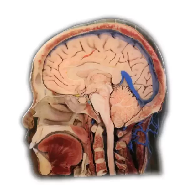
- Brain and nervous system models
- Liver, kidney, and digestive organs
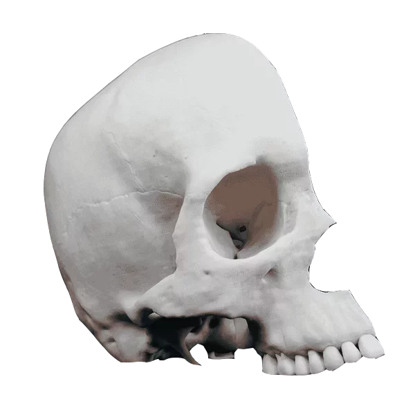
- Skeletal structures such as skulls and joints
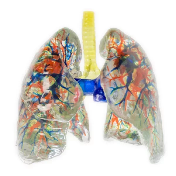
- Respiratory organs like lungs, etc.
Manufacturers can also create a 3D print human model of an entire body region. Because the models are based on digital files, they can be reproduced whenever needed.
What Are the Applications of 3D Printed Anatomical Models?
The human anatomical model improves our understanding and knowledge of various anatomical structures. Its utility spans three main areas:
1. Anatomy Teaching
The most common use of 3D printed anatomical models is in medical education. Students studying anatomy need to understand complex spatial relationships between organs, muscles, and bones. A 3D model allows them to observe these relationships directly.
Unlike real anatomical specimens, 3D printing anatomical models can be used repeatedly without degradation. They are also easier to transport and maintain. In many universities, these models now complement traditional anatomy labs and digital simulations.
Another benefit is accessibility. Schools that cannot maintain cadaver laboratories can still provide hands-on learning using 3D printed anatomical models.
2. Surgical Planning
In clinical surgery, 3D printed human models are also widely used.
For example, doctors can study the exact shape and position of organs or tumors before surgery. Surgeons may use 3D print body parts, such as a patient-specific heart or liver model. This allows them to plan incisions, evaluate surgical pathways, and reduce unexpected complications during operations.
In difficult cases, a 3D print human model of the affected area helps the surgical team practice or simulate the procedure beforehand.
3. Doctor-Patient Communication
Medical explanations can be difficult for patients to understand. Imaging scans often look confusing to people without medical training.
3D printed anatomical models make communication much easier. When doctors show a physical model of the affected organ, patients can immediately see where the problem is located.
For example, a surgeon may present a model showing a tumor or structural defect. Using 3D print body parts, physicians can explain treatment options more clearly. Patients often feel more confident when they understand their condition.
What Are the Advantages of 3D Printed Anatomical Models?
The shift towards using these models is driven by several distinct advantages:
1. High Anatomical Accuracy
Because these models are based on real medical imaging data, they reflect the subtle nuances of human biology. This includes the tiny textures of bone and the intricate pathways of blood vessels.
2. Customizability
Each 3D model of human anatomy can be customized according to specific needs.
Depending on the requirements of different interactive anatomical models, various printing effects can be selected, including full-color hard printing, soft-hard composite printing, and full-color soft printing. The model materials are respectively transparent molded materials and opaque molded materials, and the automatic mode is selected for printing support.
3. Durability
Real anatomical specimens require careful preservation. In contrast, 3D printed anatomical models are durable and easy to maintain.
3D printing anatomical models is made from strong polymers or specialized composite materials. These materials resist damage during handling and can be used repeatedly in classrooms or training sessions.
Durability also makes 3D print human model replicas practical for large teaching groups where models are frequently handled.
4. Overcoming Specimen Shortages
Many medical institutions face shortages of real anatomical specimens due to donation, ethical, and logistical limitations. This restricts hands-on training opportunities.
3D printed anatomical models help solve this issue. Schools and hospitals can purchase the required number of models based on demand and agree on delivery times with manufacturers.
This makes anatomy education more accessible worldwide.
About Digihuman 3D Printed Anatomical Models
If you are looking for 3D printed models that can rival real anatomical specimens, Digihuman offers reliable solutions.
Digihuman specializes in manufacturing 3D printed models for anatomy education. We:
- Utilize high-precision digital human datasets and select formalin-fixed cadaver specimens as references and benchmarks for the models.
- Extract voxels from the surface volume data of each anatomical structure within the original sectiondataset to generate texture maps for the geometric models. This ensures a realistic appearance.
- Use full-color and multi-material 3D printers for model production, supporting customization with singlehardness materials, soft and hard combination materials, transparent packaging materials, etc.
The final 3D printed models possess a weightiness, hardness, tactile feel, and operation experience similar to real anatomical specimens.
For any customization requirements for 3D printed anatomical models, feel free to contact us!
Conclusion
Overall, 3D printed anatomical models support anatomy teaching, assist surgical planning, and improve doctor-patient communication. With high accuracy, customization options, and long-term durability, these models are becoming an essential part of modern healthcare training.
What’s important is to choose high-quality and realistic printing models to support long-term teaching needs. If you have any requirements, quote Digihuman for the best price today!

Digihuman Technology Shines at WHX 2026 in Dubai, Delivering a Chinese Solution for Global Medical Anatomy Education!
From February 9th to 12th, the World Health Expo (WHX 2026) was held in Dubai, UAE. The event brought together...

Strengthening China–Africa Cooperation · Advancing the Future of Medical Education Senior South African Government Delegation Visits Digihuman
On January 21, 2026, a senior delegation from South Africa, including the Chair of the National Assembly Affairs Committee, the...

Digihuman Showcases at IMSH 2026, the World’s Premier Medical Simulation Event
From January 11 to 13, IMSH — the most influential annual event in the global medical simulation community — was...